1X7Y

Crystal structure of the human mitochondrial branched-chain alpha-ketoacid dehydrogenase


Experimental Data Snapshot

  • Method: X-RAY DIFFRACTION
  • Resolution: 1.57 Å
  • R-Value Free: 0.167 
  • R-Value Work: 0.150 
  • R-Value Observed: 0.150 

wwPDB Validation   3D Report Full Report


This is version 2.2 of the entry. See complete history


Literature

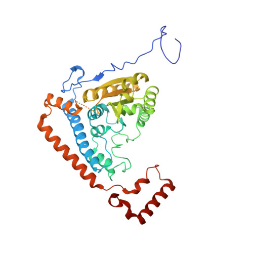
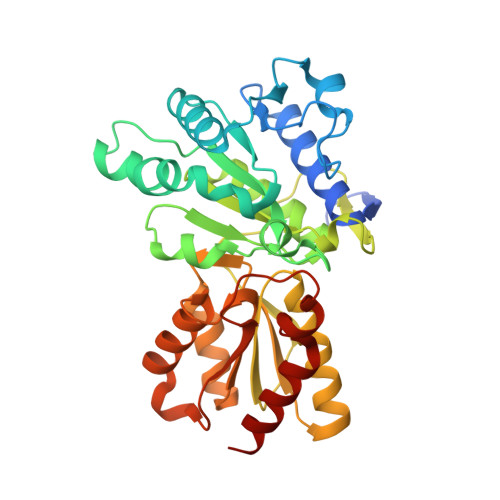
Molecular mechanism for regulation of the human mitochondrial branched-chain alpha-ketoacid dehydrogenase complex by phosphorylation

Wynn, R.M.Kato, M.Machius, M.Chuang, J.L.Li, J.Tomchick, D.R.Chuang, D.T.

(2004) Structure 12: 2185-2196

  • DOI: https://doi.org/10.1016/j.str.2004.09.013
  • Primary Citation of Related Structures:  
    1U5B, 1X7W, 1X7X, 1X7Y, 1X7Z, 1X80

  • PubMed Abstract: 

    The human mitochondrial branched-chain alpha-ketoacid dehydrogenase complex (BCKDC) is a 4 MDa macromolecular machine comprising three catalytic components (E1b, E2b, and E3), a kinase, and a phosphatase. The BCKDC overall activity is tightly regulated by phosphorylation in response to hormonal and dietary stimuli. We report that phosphorylation of Ser292-alpha in the E1b active site channel results in an order-to-disorder transition of the conserved phosphorylation loop carrying the phosphoryl serine. The conformational change is triggered by steric clashes of the phosphoryl group with invariant His291-alpha that serves as an indispensable anchor for the phosphorylation loop through bound thiamin diphosphate. Phosphorylation of Ser292-alpha does not severely impede the E1b-dependent decarboxylation of alpha-ketoacids. However, the disordered loop conformation prevents phosphorylated E1b from binding the E2b lipoyl-bearing domain, which effectively shuts off the E1b-catalyzed reductive acylation reaction and therefore completely inactivates BCKDC. This mechanism provides a paradigm for regulation of mitochondrial alpha-ketoacid dehydrogenase complexes by phosphorylation.


  • Organizational Affiliation

    Department of Biochemistry, University of Texas Southwestern Medical Center, Dallas, TX 75390, USA.


Macromolecules
Find similar proteins by:  (by identity cutoff)  |  3D Structure
Entity ID: 1
MoleculeChains Sequence LengthOrganismDetailsImage
2-oxoisovalerate dehydrogenase alpha subunit400Homo sapiensMutation(s): 1 
Gene Names: BCKDHA
EC: 1.2.4.4
UniProt & NIH Common Fund Data Resources
Find proteins for P12694 (Homo sapiens)
Explore P12694 
Go to UniProtKB:  P12694
PHAROS:  P12694
GTEx:  ENSG00000248098 
Entity Groups  
Sequence Clusters30% Identity50% Identity70% Identity90% Identity95% Identity100% Identity
UniProt GroupP12694
Sequence Annotations
Expand
  • Reference Sequence
Find similar proteins by:  (by identity cutoff)  |  3D Structure
Entity ID: 2
MoleculeChains Sequence LengthOrganismDetailsImage
2-oxoisovalerate dehydrogenase beta subunit342Homo sapiensMutation(s): 0 
Gene Names: BCKDHB
EC: 1.2.4.4
UniProt & NIH Common Fund Data Resources
Find proteins for P21953 (Homo sapiens)
Explore P21953 
Go to UniProtKB:  P21953
PHAROS:  P21953
GTEx:  ENSG00000083123 
Entity Groups  
Sequence Clusters30% Identity50% Identity70% Identity90% Identity95% Identity100% Identity
UniProt GroupP21953
Sequence Annotations
Expand
  • Reference Sequence
Small Molecules
Ligands 5 Unique
IDChains Name / Formula / InChI Key2D Diagram3D Interactions
TPP
Query on TPP

Download Ideal Coordinates CCD File 
F [auth A]THIAMINE DIPHOSPHATE
C12 H19 N4 O7 P2 S
AYEKOFBPNLCAJY-UHFFFAOYSA-O
GOL
Query on GOL

Download Ideal Coordinates CCD File 
H [auth B]GLYCEROL
C3 H8 O3
PEDCQBHIVMGVHV-UHFFFAOYSA-N
MN
Query on MN

Download Ideal Coordinates CCD File 
D [auth A]MANGANESE (II) ION
Mn
WAEMQWOKJMHJLA-UHFFFAOYSA-N
K
Query on K

Download Ideal Coordinates CCD File 
C [auth A],
G [auth B]
POTASSIUM ION
K
NPYPAHLBTDXSSS-UHFFFAOYSA-N
CL
Query on CL

Download Ideal Coordinates CCD File 
E [auth A]CHLORIDE ION
Cl
VEXZGXHMUGYJMC-UHFFFAOYSA-M
Experimental Data & Validation

Experimental Data

  • Method: X-RAY DIFFRACTION
  • Resolution: 1.57 Å
  • R-Value Free: 0.167 
  • R-Value Work: 0.150 
  • R-Value Observed: 0.150 
  • Space Group: P 31 2 1
Unit Cell:
Length ( Å )Angle ( ˚ )
a = 145.672α = 90
b = 145.672β = 90
c = 69.236γ = 120
Software Package:
Software NamePurpose
HKL-2000data collection
HKL-2000data reduction
AMoREphasing
REFMACrefinement
HKL-2000data scaling

Structure Validation

View Full Validation Report


Entry History 

Deposition Data

Revision History  (Full details and data files)

  • Version 1.0: 2004-11-23
    Type: Initial release
  • Version 1.1: 2008-04-30
    Changes: Version format compliance
  • Version 1.2: 2011-07-13
    Changes: Derived calculations, Version format compliance
  • Version 2.0: 2021-08-04
    Changes: Atomic model, Data collection, Derived calculations, Non-polymer description, Structure summary
  • Version 2.1: 2021-10-20
    Changes: Database references
  • Version 2.2: 2023-08-23
    Changes: Data collection, Refinement description