1X7D

Crystal Structure Analysis of Ornithine Cyclodeaminase Complexed with NAD and ornithine to 1.6 Angstroms


Experimental Data Snapshot

  • Method: X-RAY DIFFRACTION
  • Resolution: 1.60 Å
  • R-Value Free: 0.190 
  • R-Value Work: 0.170 
  • R-Value Observed: 0.170 

wwPDB Validation   3D Report Full Report


Ligand Structure Quality Assessment 


This is version 1.4 of the entry. See complete history


Literature

Ornithine Cyclodeaminase: Structure, Mechanism of Action, and Implications for the u-Crystallin Family

Goodman, J.L.Wang, S.Alam, S.Ruzicka, F.J.Frey, P.A.Wedekind, J.E.

(2004) Biochemistry 43: 13883-13891

  • DOI: https://doi.org/10.1021/bi048207i
  • Primary Citation of Related Structures:  
    1U7H, 1X7D

  • PubMed Abstract: 

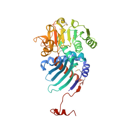
    Ornithine cyclodeaminase catalyzes the conversion of L-ornithine to L-proline by an NAD(+)-dependent hydride transfer reaction that culminates in ammonia elimination. Phylogenetic comparisons of amino acid sequences revealed that the enzyme belongs to the mu-crystallin protein family whose three-dimensional fold has not been reported. Here we describe the crystal structure of ornithine cyclodeaminase in complex with NADH, refined to 1.80 A resolution. The enzyme consists of a homodimeric fold whose subunits comprise two functional regions: (i) a novel substrate-binding domain whose antiparallel beta-strands form a 14-stranded barrel at the oligomeric interface and (ii) a canonical Rossmann fold that interacts with a single dinucleotide positioned for re hydride transfer. The adenosyl moiety of the cofactor resides in a solvent-exposed crevice on the protein surface and makes contact with a "domain-swapped"-like coil-helix module originating from the dyad-related molecule. Diffraction data were also collected to 1.60 A resolution on crystals grown in the presence of l-ornithine. The structure revealed that the substrate carboxyl group interacts with the side chains of Arg45, Lys69, and Arg112. In addition, the ammonia leaving group hydrogen bonds to the side chain of Asp228 and the site of hydride transfer is 3.8 A from C4 of the nicotinamide. The absence of an appropriately positioned water suggested that a previously proposed mechanism that calls for hydrolytic elimination of the imino intermediate must be reconsidered. A more parsimonious description of the chemical mechanism is proposed and discussed in relation to the structure and function of mu-crystallins.


  • Organizational Affiliation

    Department of Biochemistry and Biophysics, University of Rochester School of Medicine and Dentistry, Box 712, Rochester, New York 14642, USA.


Macromolecules
Find similar proteins by:  (by identity cutoff)  |  3D Structure
Entity ID: 1
MoleculeChains Sequence LengthOrganismDetailsImage
ornithine cyclodeaminase
A, B
350Pseudomonas putidaMutation(s): 8 
Gene Names: ORNITHINE CYCLASE (DEAMINATING)
EC: 4.3.1.12
UniProt
Find proteins for Q88H32 (Pseudomonas putida (strain ATCC 47054 / DSM 6125 / CFBP 8728 / NCIMB 11950 / KT2440))
Explore Q88H32 
Go to UniProtKB:  Q88H32
Entity Groups  
Sequence Clusters30% Identity50% Identity70% Identity90% Identity95% Identity100% Identity
UniProt GroupQ88H32
Sequence Annotations
Expand
  • Reference Sequence
Small Molecules
Ligands 5 Unique
IDChains Name / Formula / InChI Key2D Diagram3D Interactions
NAD
Query on NAD

Download Ideal Coordinates CCD File 
D [auth A],
L [auth B]
NICOTINAMIDE-ADENINE-DINUCLEOTIDE
C21 H27 N7 O14 P2
BAWFJGJZGIEFAR-NNYOXOHSSA-N
MES
Query on MES

Download Ideal Coordinates CCD File 
M [auth B]2-(N-MORPHOLINO)-ETHANESULFONIC ACID
C6 H13 N O4 S
SXGZJKUKBWWHRA-UHFFFAOYSA-N
ORN
Query on ORN

Download Ideal Coordinates CCD File 
E [auth A],
F [auth A]
L-ornithine
C5 H12 N2 O2
AHLPHDHHMVZTML-BYPYZUCNSA-N
MPD
Query on MPD

Download Ideal Coordinates CCD File 
G [auth A]
H [auth A]
I [auth A]
J [auth A]
N [auth B]
G [auth A],
H [auth A],
I [auth A],
J [auth A],
N [auth B],
O [auth B]
(4S)-2-METHYL-2,4-PENTANEDIOL
C6 H14 O2
SVTBMSDMJJWYQN-YFKPBYRVSA-N
NA
Query on NA

Download Ideal Coordinates CCD File 
C [auth A],
K [auth B]
SODIUM ION
Na
FKNQFGJONOIPTF-UHFFFAOYSA-N
Modified Residues  1 Unique
IDChains TypeFormula2D DiagramParent
MSE
Query on MSE
A, B
L-PEPTIDE LINKINGC5 H11 N O2 SeMET
Experimental Data & Validation

Experimental Data

  • Method: X-RAY DIFFRACTION
  • Resolution: 1.60 Å
  • R-Value Free: 0.190 
  • R-Value Work: 0.170 
  • R-Value Observed: 0.170 
  • Space Group: P 21 21 21
Unit Cell:
Length ( Å )Angle ( ˚ )
a = 69.88α = 90
b = 78.62β = 90
c = 119.94γ = 90
Software Package:
Software NamePurpose
CNSrefinement
ADSCdata collection
CrystalCleardata scaling
CNSphasing

Structure Validation

View Full Validation Report


Ligand Structure Quality Assessment 


Entry History 

Deposition Data

Revision History  (Full details and data files)

  • Version 1.0: 2004-11-09
    Type: Initial release
  • Version 1.1: 2008-04-30
    Changes: Version format compliance
  • Version 1.2: 2011-07-13
    Changes: Non-polymer description, Version format compliance
  • Version 1.3: 2023-08-23
    Changes: Data collection, Database references, Derived calculations, Refinement description
  • Version 1.4: 2023-11-15
    Changes: Data collection