1X0I

Crystal Structure of the Acid Blue Form of Bacteriorhodopsin


Experimental Data Snapshot

  • Method: X-RAY DIFFRACTION
  • Resolution: 2.30 Å
  • R-Value Free: 0.282 
  • R-Value Work: 0.238 
  • R-Value Observed: 0.238 

wwPDB Validation   3D Report Full Report


Ligand Structure Quality Assessment 


This is version 1.4 of the entry. See complete history


Literature

Crystal Structures of Acid Blue and Alkaline Purple Forms of Bacteriorhodopsin

Okumura, H.Murakami, M.Kouyama, T.

(2005) J Mol Biol 351: 481-495

  • DOI: https://doi.org/10.1016/j.jmb.2005.06.026
  • Primary Citation of Related Structures:  
    1X0I

  • PubMed Abstract: 

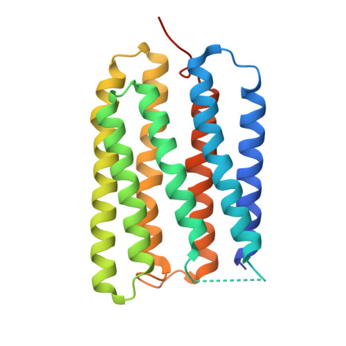
    Bacteriorhodopsin, a light-driven proton pump found in the purple membrane of Halobacterium salinarum, exhibits purple at neutral pH but its color is sensitive to pH. Here, structures are reported for an acid blue form and an alkaline purple form of wild-type bacteriorhodopsin. When the P622 crystal prepared at pH 5.2 was acidified with sulfuric acid, its color turned to blue with a pKa of 3.5 and a Hill coefficient of 2. Diffraction data at pH 2-5 indicated that the purple-to-blue transition accompanies a large structural change in the proton release channel; i.e. the extracellular half of helix C moves towards helix G, narrowing the proton release channel and expelling a water molecule from a micro-cavity in the vicinity of the retinal Schiff base. In this respect, the acid-induced structural change resembles the structural change observed upon formation of the M intermediate. But, the acid blue form contains a sulfate ion in a site(s) near Arg82 that is created by re-orientations of the carboxyl groups of Glu194 and Glu204, residues comprising the proton release complex. This result suggests that proton uptake by the proton release complex evokes the anion binding, which in turn induces protonation of Asp85, a key residue regulating the absorption spectrum of the chromophore. Interestingly, a pronounced structural change in the proton release complex was also observed at high pH; i.e. re-orientation of Glu194 towards Tyr83 was found to take place at around pH 10. This alkaline transition is suggested to be accompanied by proton release from the proton release complex and responsible for rapid formation of the M intermediate at high pH.


  • Organizational Affiliation

    Department of Physics, Graduate School of Science, Nagoya University, Nagoya 464-8602, Japan.


Macromolecules
Find similar proteins by:  (by identity cutoff)  |  3D Structure
Entity ID: 1
MoleculeChains Sequence LengthOrganismDetailsImage
BacteriorhodopsinA [auth 1]248Halobacterium salinarumMutation(s): 0 
Membrane Entity: Yes 
UniProt
Find proteins for P02945 (Halobacterium salinarum (strain ATCC 700922 / JCM 11081 / NRC-1))
Explore P02945 
Go to UniProtKB:  P02945
Entity Groups  
Sequence Clusters30% Identity50% Identity70% Identity90% Identity95% Identity100% Identity
UniProt GroupP02945
Sequence Annotations
Expand
  • Reference Sequence
Small Molecules
Ligands 4 Unique
IDChains Name / Formula / InChI Key2D Diagram3D Interactions
L3P
Query on L3P

Download Ideal Coordinates CCD File 
D [auth 1],
F [auth 1],
G [auth 1]
2,3-DI-O-PHYTANLY-3-SN-GLYCERO-1-PHOSPHORYL-3'-SN-GLYCEROL-1'-PHOSPHATE
C46 H94 O11 P2
TZXJQSKPTCRGCA-VZSPAKCESA-L
L2P
Query on L2P

Download Ideal Coordinates CCD File 
E [auth 1]2,3-DI-PHYTANYL-GLYCEROL
C43 H88 O3
ISDBCJSGCHUHFI-UMZPFTBHSA-N
RET
Query on RET

Download Ideal Coordinates CCD File 
C [auth 1]RETINAL
C20 H28 O
NCYCYZXNIZJOKI-OVSJKPMPSA-N
SO4
Query on SO4

Download Ideal Coordinates CCD File 
B [auth 1]SULFATE ION
O4 S
QAOWNCQODCNURD-UHFFFAOYSA-L
Experimental Data & Validation

Experimental Data

  • Method: X-RAY DIFFRACTION
  • Resolution: 2.30 Å
  • R-Value Free: 0.282 
  • R-Value Work: 0.238 
  • R-Value Observed: 0.238 
  • Space Group: P 6 2 2
Unit Cell:
Length ( Å )Angle ( ˚ )
a = 102.64α = 90
b = 102.64β = 90
c = 109.04γ = 120
Software Package:
Software NamePurpose
CNSrefinement
MOSFLMdata reduction
CCP4data scaling
XTALVIEWrefinement

Structure Validation

View Full Validation Report


Ligand Structure Quality Assessment 


Entry History 

Deposition Data

Revision History  (Full details and data files)

  • Version 1.0: 2005-08-02
    Type: Initial release
  • Version 1.1: 2008-04-30
    Changes: Version format compliance
  • Version 1.2: 2011-07-13
    Changes: Version format compliance
  • Version 1.3: 2015-09-09
    Changes: Other
  • Version 1.4: 2023-10-25
    Changes: Data collection, Database references, Derived calculations, Refinement description