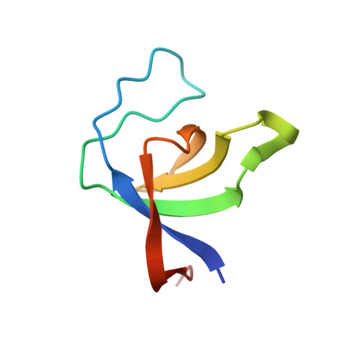
The 1.1 A resolution crystal structure of the p130cas SH3 domain and ramifications for ligand selectivity
Wisniewska, M., Bossenmaier, B., Georges, G., Hesse, F., Dangl, M., Kunkele, K.P., Ioannidis, I., Huber, R., Engh, R.A.(2005) J Mol Biol 347: 1005-1014
- PubMed: 15784259
- DOI: https://doi.org/10.1016/j.jmb.2005.02.017
- Primary Citation of Related Structures:
1WYX - PubMed Abstract:
The Crk-associated tyrosine kinase substrate p130cas (CAS) is a docking protein containing an SH3 domain near its N terminus, followed by a short proline-rich segment, a large central substrate domain composed of 15 repeats of the four amino acid sequence YxxP, a serine-rich region and a carboxy-terminal domain, which possesses consensus binding sites for the SH2 and SH3 domains of Src (YDYV and RPLPSPP, respectively). The SH3 domain of CAS mediates its interaction with several proteins involved in signaling pathways such as focal adhesion kinase (FAK), tyrosine phosphatases PTP1B and PTP-PEST, and the guanine nucleotide exchange factor C3G. As a homolog of the corresponding Src docking domain, the CAS SH3 domain binds to proline-rich sequences (PxxP) of its interacting partners that can adopt a polyproline type II helix. We have determined a high-resolution X-ray structure of the recombinant human CAS SH3 domain. The domain, residues 1-69, crystallized in two related space groups, P2(1) and C222(1), that provided diffraction data to 1.1 A and 2.1 A, respectively. The crystal structure shows, in addition to the conserved SH3 domain architecture, the way in which the CAS characteristic amino acids form an atypically charged ligand-binding surface. This arrangement provides a rationale for the unusual ligand recognition motif exhibited by the CAS SH3 domain. The structure enables modelling of the docking interactions to its ligands, for example from focal adhesion kinase, and supports structure-based drug design of inhibitors of the CAS-FAK interaction.
Organizational Affiliation:
Max Planck Institut für Biochemie, Strukturforschung, D-82152 Martinsried, Germany.