1WXX

Crystal structure of Tt1595, a putative SAM-dependent methyltransferase from Thermus thermophillus HB8


Experimental Data Snapshot

  • Method: X-RAY DIFFRACTION
  • Resolution: 1.80 Å
  • R-Value Free: 0.229 
  • R-Value Work: 0.203 
  • R-Value Observed: 0.203 

wwPDB Validation   3D Report Full Report


This is version 1.4 of the entry. See complete history


Literature

Structures of a putative RNA 5-methyluridine methyltransferase, Thermus thermophilus TTHA1280, and its complex with S-adenosyl-L-homocysteine.

Pioszak, A.A.Murayama, K.Nakagawa, N.Ebihara, A.Kuramitsu, S.Shirouzu, M.Yokoyama, S.

(2005) Acta Crystallogr Sect F Struct Biol Cryst Commun 61: 867-874

  • DOI: https://doi.org/10.1107/S1744309105029842
  • Primary Citation of Related Structures:  
    1WXW, 1WXX, 2CWW

  • PubMed Abstract: 

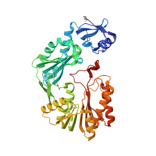
    The Thermus thermophilus hypothetical protein TTHA1280 belongs to a family of predicted S-adenosyl-L-methionine (AdoMet) dependent RNA methyltransferases (MTases) present in many bacterial and archaeal species. Inspection of amino-acid sequence motifs common to class I Rossmann-fold-like MTases suggested a specific role as an RNA 5-methyluridine MTase. Selenomethionine (SeMet) labelled and native versions of the protein were expressed, purified and crystallized. Two crystal forms of the SeMet-labelled apoprotein were obtained: SeMet-ApoI and SeMet-ApoII. Cocrystallization of the native protein with S-adenosyl-L-homocysteine (AdoHcy) yielded a third crystal form, Native-AdoHcy. The SeMet-ApoI structure was solved by the multiple anomalous dispersion method and refined at 2.55 A resolution. The SeMet-ApoII and Native-AdoHcy structures were solved by molecular replacement and refined at 1.80 and 2.60 A, respectively. TTHA1280 formed a homodimer in the crystals and in solution. Each subunit folds into a three-domain structure composed of a small N-terminal PUA domain, a central alpha/beta-domain and a C-terminal Rossmann-fold-like MTase domain. The three domains form an overall clamp-like shape, with the putative active site facing a deep cleft. The architecture of the active site is consistent with specific recognition of uridine and catalysis of methyl transfer to the 5-carbon position. The cleft is suitable in size and charge distribution for binding single-stranded RNA.


  • Organizational Affiliation

    RIKEN Genomic Sciences Center, Yokohama, Japan.


Macromolecules
Find similar proteins by:  (by identity cutoff)  |  3D Structure
Entity ID: 1
MoleculeChains Sequence LengthOrganismDetailsImage
hypothetical protein TTHA1280
A, B, C, D
382Thermus thermophilus HB8Mutation(s): 4 
UniProt
Find proteins for Q5SIT4 (Thermus thermophilus (strain ATCC 27634 / DSM 579 / HB8))
Explore Q5SIT4 
Go to UniProtKB:  Q5SIT4
Entity Groups  
Sequence Clusters30% Identity50% Identity70% Identity90% Identity95% Identity100% Identity
UniProt GroupQ5SIT4
Sequence Annotations
Expand
  • Reference Sequence
Small Molecules
Ligands 2 Unique
IDChains Name / Formula / InChI Key2D Diagram3D Interactions
PO4
Query on PO4

Download Ideal Coordinates CCD File 
F [auth A]
G [auth A]
H [auth A]
I [auth A]
J [auth A]
F [auth A],
G [auth A],
H [auth A],
I [auth A],
J [auth A],
L [auth B],
M [auth B],
O [auth C]
PHOSPHATE ION
O4 P
NBIIXXVUZAFLBC-UHFFFAOYSA-K
K
Query on K

Download Ideal Coordinates CCD File 
E [auth A],
K [auth B],
N [auth C],
P [auth D]
POTASSIUM ION
K
NPYPAHLBTDXSSS-UHFFFAOYSA-N
Modified Residues  1 Unique
IDChains TypeFormula2D DiagramParent
MSE
Query on MSE
A, B, C, D
L-PEPTIDE LINKINGC5 H11 N O2 SeMET
Experimental Data & Validation

Experimental Data

  • Method: X-RAY DIFFRACTION
  • Resolution: 1.80 Å
  • R-Value Free: 0.229 
  • R-Value Work: 0.203 
  • R-Value Observed: 0.203 
  • Space Group: P 1
Unit Cell:
Length ( Å )Angle ( ˚ )
a = 77.551α = 96.34
b = 82.318β = 90.14
c = 79.616γ = 103.06
Software Package:
Software NamePurpose
CNSrefinement
HKL-2000data reduction
SCALEPACKdata scaling
MOLREPphasing

Structure Validation

View Full Validation Report


Entry History 

Revision History  (Full details and data files)

  • Version 1.0: 2005-08-02
    Type: Initial release
  • Version 1.1: 2008-04-30
    Changes: Version format compliance
  • Version 1.2: 2011-07-13
    Changes: Source and taxonomy, Version format compliance
  • Version 1.3: 2023-10-25
    Changes: Data collection, Database references, Derived calculations, Refinement description
  • Version 1.4: 2023-11-15
    Changes: Data collection