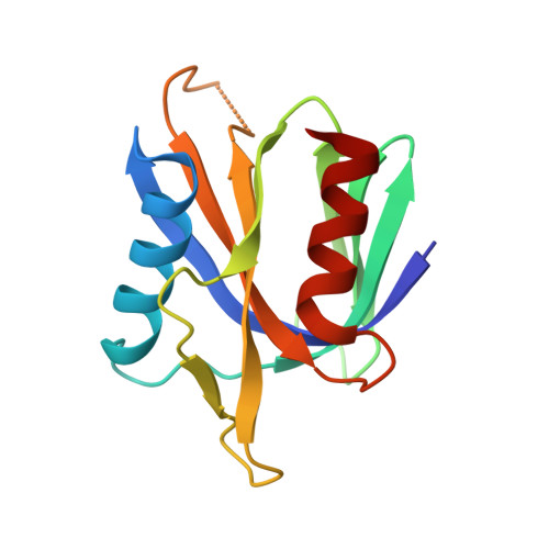
1WVH

Crystal structure of tensin1 PTB domain


Experimental Data Snapshot

  • Method: X-RAY DIFFRACTION
  • Resolution: 1.50 Å
  • R-Value Free: 0.236 
  • R-Value Work: 0.218 
  • R-Value Observed: 0.218 

wwPDB Validation   3D Report Full Report


This is version 1.3 of the entry. See complete history


Literature

Structure of the PTB domain of tensin1 and a model for its recruitment to fibrillar adhesions.

McCleverty, C.J.Lin, D.C.Liddington, R.C.

(2007) Protein Sci 16: 1223-1229

  • DOI: https://doi.org/10.1110/ps.072798707
  • Primary Citation of Related Structures:  
    1WVH

  • PubMed Abstract: 

    Tensin is a cytoskeletal protein that links integrins to the actin cytoskeleton at sites of cell-matrix adhesion. Here we describe the crystal structure of the phosphotyrosine-binding (PTB) domain of tensin1, and show that it binds integrins in an NPxY-dependent fashion. Alanine mutagenesis of both the PTB domain and integrin tails supports a model of integrin binding similar to that of the PTB-like domain of talin. However, we also show that phosphorylation of the NPxY tyrosine, which disrupts talin binding, has a negligible effect on tensin binding. This suggests that tyrosine phosphorylation of integrin, which occurs during the maturation of focal adhesions, could act as a switch to promote the migration of tensin-integrin complexes into fibronectin-mediated fibrillar adhesions.


  • Organizational Affiliation

    Cancer Center, Burnham Institute for Medical Research, La Jolla, California 92037, USA.


Macromolecules
Find similar proteins by:  (by identity cutoff)  |  3D Structure
Entity ID: 1
MoleculeChains Sequence LengthOrganismDetailsImage
Tensin134Gallus gallusMutation(s): 0 
UniProt
Find proteins for Q04205 (Gallus gallus)
Explore Q04205 
Go to UniProtKB:  Q04205
Entity Groups  
Sequence Clusters30% Identity50% Identity70% Identity90% Identity95% Identity100% Identity
UniProt GroupQ04205
Sequence Annotations
Expand
  • Reference Sequence
Experimental Data & Validation

Experimental Data

  • Method: X-RAY DIFFRACTION
  • Resolution: 1.50 Å
  • R-Value Free: 0.236 
  • R-Value Work: 0.218 
  • R-Value Observed: 0.218 
  • Space Group: I 4
Unit Cell:
Length ( Å )Angle ( ˚ )
a = 75.68α = 90
b = 75.68β = 90
c = 48.69γ = 90
Software Package:
Software NamePurpose
DENZOdata reduction
SCALEPACKdata scaling
SOLVEphasing
CNSrefinement

Structure Validation

View Full Validation Report


Entry History 

Deposition Data

Revision History  (Full details and data files)

  • Version 1.0: 2005-12-13
    Type: Initial release
  • Version 1.1: 2008-04-30
    Changes: Version format compliance
  • Version 1.2: 2011-07-13
    Changes: Version format compliance
  • Version 1.3: 2024-03-13
    Changes: Data collection, Database references