Kinetic and structural analysis of alpha-D-Glucose-1-phosphate cytidylyltransferase from Salmonella typhi.
Koropatkin, N.M., Cleland, W.W., Holden, H.M.(2005) J Biol Chem 280: 10774-10780
- PubMed: 15634670
- DOI: https://doi.org/10.1074/jbc.M414111200
- Primary Citation of Related Structures:
1WVC - PubMed Abstract:
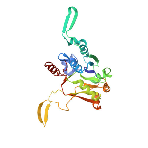
Tyvelose is a 3,6-dideoxyhexose found in the O-antigen of the surface lipopolysaccharides of some pathogenic bacteria. It is synthesized via a complex biochemical pathway that is initiated by the formation of CDP-D-glucose. The production of this ligand is catalyzed by the enzyme glucose-1-phosphate cytidylyltransferase, which utilizes alpha-D-glucose 1-phosphate and MgCTP as substrates. Previous x-ray crystallographic investigations have demonstrated that the Salmonella typhi enzyme complexed with the product CDP-glucose is a fully integrated hexamer displaying 32 point group symmetry. The binding pocket for CDP-glucose is shared between two subunits. Here we describe both a detailed kinetic analysis of the cytidylyltransferase and a structural investigation of the enzyme complexed with MgCTP. These data demonstrate that the reaction catalyzed by the cytidylyltransferase proceeds via a sequential rather than a Bi Bi ping-pong mechanism as was previously reported. Additionally, the enzyme utilizes both CTP and UTP equally well as substrates. The structure of the enzyme with bound MgCTP reveals that the binding pocket for the nucleotide is contained within one subunit rather than shared between two. Key side chains involved in nucleotide binding include Thr(14), Arg(15), Lys(25), and Arg(111). In the previous structure of the enzyme complexed with CDP-glucose, those residues defined by Thr(14) to Ile(21) were disordered. The kinetic and x-ray crystallographic data presented here support a mechanism for this enzyme that is similar to that reported for the glucose-1-phosphate thymidylyltransferases.
Organizational Affiliation:
Department of Biochemistry, University of Wisconsin, Madison, Wisconsin 53706, USA.