Structure of the Bacillus subtilis phage SPO1-encoded type II DNA-binding protein TF1 in solution.
Jia, X., Grove, A., Ivancic, M., Hsu, V.L., Geiduscheck, E.P., Kearns, D.R.(1996) J Mol Biol 263: 259-268
- PubMed: 8913305
- DOI: https://doi.org/10.1006/jmbi.1996.0573
- Primary Citation of Related Structures:
1WTU - PubMed Abstract:
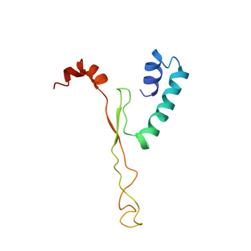
The solution structure of a type II DNA-binding protein, the bacteriophage SPO1-encoded transcription factor 1 (TF1), was determined using NMR spectroscopy. Selective 2H-labeling, 13C-labeling and isotopic heterodimers were used to distinguish contacts between and within monomers of the dimeric protein. A total of 1914 distance and dihedral angle constraints derived from NMR experiments were used in structure calculations using restrained molecular dynamics and simulated annealing protocols. The ensemble of 30 calculated structures has a root-mean-square deviation (r.m.s.d.) of 0.9 A, about the average structure for the backbone atoms, and 1.2 A for all heavy-atoms of the dimeric core (helices 1 and 2) and the beta-sheets. A severe helix distortion at residues 92-93 in the middle of helix 3 is associated with r.m.s.d. of approximately 1.5 A for the helix 3 backbone. Deviations of approximately 5 A or larger are noted for the very flexible beta-ribbon arms that constitute part of a proposed DNA-binding region. A structural model of TF1 has been calculated based on the previously reported crystal structure of the homologous HU protein and this model was used as the starting structure for calculations. A comparison between the calculated average solution structure of TF1 and a solution structure of HU indicates a similarity in the dimeric core (excluding the nine amino acid residue tail) with pairwise deviations of 2 to 3 A. The largest deviations between the average structure and the HU solution structure were found in the beta-ribbon arms, as expected. A 4 A deviation is found at residue 15 of TF1 which is in a loop connecting two helical segments; it has been reported that substitution of Glu15 by Gly increases the thermostability of TF1. The homology between TF1 and other proteins of this family leads us to anticipate similar tertiary structures.
Organizational Affiliation:
Department of Chemistry and Biochemistry, University of California, San Diego, La Jolla 92093, USA.