1WRA

Crystal Structure of Phosphorylcholine Esterase Domain of the Virulence Factor Choline Binding Protein E from Streptococcus Pneumoniae


Experimental Data Snapshot

  • Method: X-RAY DIFFRACTION
  • Resolution: 2.00 Å
  • R-Value Free: 0.204 
  • R-Value Work: 0.174 
  • R-Value Observed: 0.176 

wwPDB Validation   3D Report Full Report


Ligand Structure Quality Assessment 


This is version 1.3 of the entry. See complete history


Literature

Crystal structure of phosphorylcholine esterase domain of the virulence factor choline-binding protein e from streptococcus pneumoniae: new structural features among the metallo-beta-lactamase superfamily

Garau, G.Lemaire, D.Vernet, T.Dideberg, O.Di Guilmi, A.M.

(2005) J Biol Chem 280: 28591-28600

  • DOI: https://doi.org/10.1074/jbc.M502744200
  • Primary Citation of Related Structures:  
    1WRA

  • PubMed Abstract: 

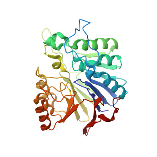
    Streptococcus pneumoniae is the worldwide leading cause of deaths from invasive infections such as pneumoniae, sepsis, and meningitidis in children and the elderly. Nasopharyngeal colonization, which plays a key role in the development of pneumococcal disease, is highly dependent on a family of surface-exposed proteins, the choline-binding proteins (CBPs). Here we report the crystal structure of phosphorylcholine esterase (Pce), the catalytic domain of choline-binding protein E (CBPE), which has been shown to be crucial for host/pathogen interaction processes. The unexpected features of the Pce active site reveal that this enzyme is unique among the large family of hydrolases harboring the metallo-beta-lactamase fold. The orientation and calcium stabilization features of an elongated loop, which lies on top of the active site, suggest that the cleft may be rearranged. Furthermore, the structure of Pce complexed with phosphorylcholine, together with the characterization of the enzymatic role played by two iron ions located in the active site allow us to propose a reaction mechanism reminiscent of that of purple acid phosphatase. This mechanism is supported by site-directed mutagenesis experiments. Finally, the interactions of the choline binding domain and the Pce region of CBPE with chains of teichoic acids have been modeled. The ensemble of our biochemical and structural results provide an initial understanding of the function of CBPE.


  • Organizational Affiliation

    Laboratoire de Cristallographie Macromoléculaire, Institut de Biologie Structurale Jean-Pierre Ebel (CEA-CNRS UMR 5075-UJF), 41 Rue Jules Horowitz 38027, Grenoble Cedex 1, France.


Macromolecules
Find similar proteins by:  (by identity cutoff)  |  3D Structure
Entity ID: 1
MoleculeChains Sequence LengthOrganismDetailsImage
Teichoic acid phosphorylcholine esterase/choline binding protein E (cbpE)
A, B
308Streptococcus pneumoniae R6Mutation(s): 0 
EC: 3.1.4.38
UniProt
Find proteins for Q8DQ62 (Streptococcus pneumoniae (strain ATCC BAA-255 / R6))
Explore Q8DQ62 
Go to UniProtKB:  Q8DQ62
Entity Groups  
Sequence Clusters30% Identity50% Identity70% Identity90% Identity95% Identity100% Identity
UniProt GroupQ8DQ62
Sequence Annotations
Expand
  • Reference Sequence
Small Molecules
Ligands 4 Unique
IDChains Name / Formula / InChI Key2D Diagram3D Interactions
PC
Query on PC

Download Ideal Coordinates CCD File 
G [auth A],
R [auth B]
PHOSPHOCHOLINE
C5 H15 N O4 P
YHHSONZFOIEMCP-UHFFFAOYSA-O
MPD
Query on MPD

Download Ideal Coordinates CCD File 
H [auth A]
I [auth A]
J [auth A]
K [auth A]
L [auth A]
H [auth A],
I [auth A],
J [auth A],
K [auth A],
L [auth A],
M [auth A],
S [auth B],
T [auth B],
U [auth B],
V [auth B],
W [auth B]
(4S)-2-METHYL-2,4-PENTANEDIOL
C6 H14 O2
SVTBMSDMJJWYQN-YFKPBYRVSA-N
FE
Query on FE

Download Ideal Coordinates CCD File 
C [auth A],
D [auth A],
N [auth B],
O [auth B]
FE (III) ION
Fe
VTLYFUHAOXGGBS-UHFFFAOYSA-N
CA
Query on CA

Download Ideal Coordinates CCD File 
E [auth A],
F [auth A],
P [auth B],
Q [auth B]
CALCIUM ION
Ca
BHPQYMZQTOCNFJ-UHFFFAOYSA-N
Experimental Data & Validation

Experimental Data

  • Method: X-RAY DIFFRACTION
  • Resolution: 2.00 Å
  • R-Value Free: 0.204 
  • R-Value Work: 0.174 
  • R-Value Observed: 0.176 
  • Space Group: P 41 21 2
Unit Cell:
Length ( Å )Angle ( ˚ )
a = 98.302α = 90
b = 98.302β = 90
c = 172.6γ = 90
Software Package:
Software NamePurpose
MOSFLMdata reduction
SCALAdata scaling
CCP4model building
REFMACrefinement
CCP4data scaling
CCP4phasing

Structure Validation

View Full Validation Report


Ligand Structure Quality Assessment 


Entry History 

Deposition Data

Revision History  (Full details and data files)

  • Version 1.0: 2005-05-31
    Type: Initial release
  • Version 1.1: 2008-04-30
    Changes: Version format compliance
  • Version 1.2: 2011-07-13
    Changes: Version format compliance
  • Version 1.3: 2024-03-13
    Changes: Data collection, Database references, Derived calculations, Refinement description