1WQ9

Crystal structure of VR-1, a VEGF-F from a snake venom


Experimental Data Snapshot

  • Method: X-RAY DIFFRACTION
  • Resolution: 2.00 Å
  • R-Value Free: 0.252 
  • R-Value Work: 0.206 
  • R-Value Observed: 0.206 

wwPDB Validation   3D Report Full Report


This is version 2.0 of the entry. See complete history


Literature

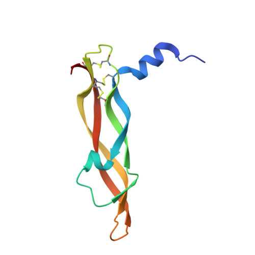
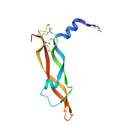
Crystal structures of novel vascular endothelial growth factors (VEGF) from snake venoms: insight into selective VEGF binding to kinase insert domain-containing receptor but not to fms-like tyrosine kinase-1.

Suto, K.Yamazaki, Y.Morita, T.Mizuno, H.

(2005) J Biol Chem 280: 2126-2131

  • DOI: https://doi.org/10.1074/jbc.M411395200
  • Primary Citation of Related Structures:  
    1WQ8, 1WQ9

  • PubMed Abstract: 

    Vascular endothelial growth factor-A (VEGF-A(165)) exerts multiple effects upon binding to the fms-like tyrosine kinase-1 (Flt-1) and the kinase insert domain-containing receptor (KDR). We recently identified two novel snake venom VEGFs (vammin and VR-1) having unique properties. These VEGFs, designated VEGF-Fs, are highly specific ligands for the kinase insert domain-containing receptor and exhibit potent biological activity both in vitro and in vivo when compared with VEGF-A(165). Here, we solved the crystal structures of vammin and VR-1 at 1.9 and 2.0 A resolutions, respectively. Both structures are very similar to each other, and these structures exhibit similar but significantly different features from the known structures of other VEGFs. These differences include a conformational difference in receptor-binding loop 3 caused by an amino acid residue insertion and a difference in surface potential on the possible binding surface for domain 3 of the receptor. These structural differences may be related to the highly selective ligand properties of VEGF-F.


  • Organizational Affiliation

    Department of Biochemistry, National Institute of Agrobiological Sciences, 2-1-2, Kannondai, Tsukuba, Ibaraki 305-8602, Japan.


Macromolecules
Find similar proteins by:  (by identity cutoff)  |  3D Structure
Entity ID: 1
MoleculeChains Sequence LengthOrganismDetailsImage
Vascular endothelial growth factor96Daboia russelii russeliiMutation(s): 0 
UniProt
Find proteins for P67861 (Daboia russelii)
Explore P67861 
Go to UniProtKB:  P67861
Entity Groups  
Sequence Clusters30% Identity50% Identity70% Identity90% Identity95% Identity100% Identity
UniProt GroupP67861
Sequence Annotations
Expand
  • Reference Sequence
Find similar proteins by:  (by identity cutoff)  |  3D Structure
Entity ID: 2
MoleculeChains Sequence LengthOrganismDetailsImage
Vascular endothelial growth factor96Daboia russelii russeliiMutation(s): 0 
UniProt
Find proteins for P67861 (Daboia russelii)
Explore P67861 
Go to UniProtKB:  P67861
Entity Groups  
Sequence Clusters30% Identity50% Identity70% Identity90% Identity95% Identity100% Identity
UniProt GroupP67861
Sequence Annotations
Expand
  • Reference Sequence
Small Molecules
Modified Residues  1 Unique
IDChains TypeFormula2D DiagramParent
PCA
Query on PCA
B
L-PEPTIDE LINKINGC5 H7 N O3GLN
Experimental Data & Validation

Experimental Data

  • Method: X-RAY DIFFRACTION
  • Resolution: 2.00 Å
  • R-Value Free: 0.252 
  • R-Value Work: 0.206 
  • R-Value Observed: 0.206 
  • Space Group: P 21 21 2
Unit Cell:
Length ( Å )Angle ( ˚ )
a = 90.348α = 90
b = 48.961β = 90
c = 54.214γ = 90
Software Package:
Software NamePurpose
CNSrefinement
HKL-2000data reduction
SCALEPACKdata scaling
CNSphasing
SHARPphasing

Structure Validation

View Full Validation Report


Entry History 

Deposition Data

Revision History  (Full details and data files)

  • Version 1.0: 2004-12-07
    Type: Initial release
  • Version 1.1: 2008-04-30
    Changes: Version format compliance
  • Version 1.2: 2011-07-13
    Changes: Version format compliance
  • Version 2.0: 2019-12-25
    Changes: Derived calculations, Polymer sequence