1WPX

Crystal structure of carboxypeptidase Y inhibitor complexed with the cognate proteinase


Experimental Data Snapshot

  • Method: X-RAY DIFFRACTION
  • Resolution: 2.70 Å
  • R-Value Free: 0.225 
  • R-Value Work: 0.183 
  • R-Value Observed: 0.183 

wwPDB Validation   3D Report Full Report


Ligand Structure Quality Assessment 


This is version 2.0 of the entry. See complete history


Literature

Structure of the carboxypeptidase y inhibitor i(c) in complex with the cognate proteinase reveals a novel mode of the proteinase-protein inhibitor interaction

Mima, J.Hayashida, M.Fujii, T.Narita, Y.Hayashi, R.Ueda, M.Hata, Y.

(2005) J Mol Biol 346: 1323-1334

  • DOI: https://doi.org/10.1016/j.jmb.2004.12.051
  • Primary Citation of Related Structures:  
    1WPX

  • PubMed Abstract: 

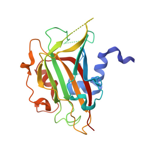
    Carboxypeptidase Y (CPY) inhibitor, IC, shows no homology to any other known proteinase inhibitors and rather belongs to the phosphatidylethanolamine-binding protein (PEBP) family. We report here on the crystal structure of the IC-CPY complex at 2.7 A resolution. The structure of IC in the complex with CPY consists of one major beta-type domain and a N-terminal helical segment. The structure of the complex contains two binding sites of IC toward CPY, the N-terminal inhibitory reactive site (the primary CPY-binding site) and the secondary CPY-binding site, which interact with the S1 substrate-binding site of CPY and the hydrophobic surface flanked by the active site of the enzyme, respectively. It was also revealed that IC had the ligand-binding site, which is conserved among PEBPs and the putative binding site of the polar head group of phospholipid. The complex structure and analyses of IC mutants for inhibitory activity and the binding to CPY demonstrate that the N-terminal inhibitory reactive site is essential both for inhibitory function and the complex formation with CPY and that the binding of IC to CPY constitutes a novel mode of the proteinase-protein inhibitor interaction. The unique binding mode of IC toward the cognate proteinase provides insights into the inhibitory mechanism of PEBPs toward serine proteinases and into the specific biological functions of IC belonging to the PEBP family as well.


  • Organizational Affiliation

    Division of Applied Life Sciences, Graduate School of Agriculture, Kyoto University, Kyoto 606-8502, Japan.


Macromolecules
Find similar proteins by:  (by identity cutoff)  |  3D Structure
Entity ID: 1
MoleculeChains Sequence LengthOrganismDetailsImage
Carboxypeptidase Y421Saccharomyces cerevisiaeMutation(s): 0 
EC: 3.4.16.5
UniProt
Find proteins for P00729 (Saccharomyces cerevisiae (strain ATCC 204508 / S288c))
Explore P00729 
Go to UniProtKB:  P00729
Entity Groups  
Sequence Clusters30% Identity50% Identity70% Identity90% Identity95% Identity100% Identity
UniProt GroupP00729
Sequence Annotations
Expand
  • Reference Sequence
Find similar proteins by:  (by identity cutoff)  |  3D Structure
Entity ID: 2
MoleculeChains Sequence LengthOrganismDetailsImage
Carboxypeptidase Y inhibitor220Saccharomyces cerevisiaeMutation(s): 0 
UniProt
Find proteins for P14306 (Saccharomyces cerevisiae (strain ATCC 204508 / S288c))
Explore P14306 
Go to UniProtKB:  P14306
Entity Groups  
Sequence Clusters30% Identity50% Identity70% Identity90% Identity95% Identity100% Identity
UniProt GroupP14306
Sequence Annotations
Expand
  • Reference Sequence
Experimental Data & Validation

Experimental Data

  • Method: X-RAY DIFFRACTION
  • Resolution: 2.70 Å
  • R-Value Free: 0.225 
  • R-Value Work: 0.183 
  • R-Value Observed: 0.183 
  • Space Group: P 21 21 21
Unit Cell:
Length ( Å )Angle ( ˚ )
a = 81.129α = 90
b = 186.59β = 90
c = 65.14γ = 90
Software Package:
Software NamePurpose
CNSrefinement
HKL-2000data reduction
SCALEPACKdata scaling
MOLREPphasing

Structure Validation

View Full Validation Report


Ligand Structure Quality Assessment 


Entry History 

Deposition Data

Revision History  (Full details and data files)

  • Version 1.0: 2005-03-01
    Type: Initial release
  • Version 1.1: 2008-04-30
    Changes: Version format compliance
  • Version 1.2: 2011-07-13
    Changes: Non-polymer description, Version format compliance
  • Version 2.0: 2020-07-29
    Type: Remediation
    Reason: Carbohydrate remediation
    Changes: Advisory, Atomic model, Data collection, Derived calculations, Non-polymer description, Structure summary