


Crystal structure of methylglyoxal synthase from Thermus thermophilus HB8
Sugahara, M., Kunishima, N.To be published.
Experimental Data Snapshot
wwPDB Validation 3D Report Full Report

Entity ID: 1 | |||||
|---|---|---|---|---|---|
| Molecule | Chains | Sequence Length | Organism | Details | Image |
| methylglyoxal synthase | 126 | Thermus thermophilus HB8 | Mutation(s): 0 EC: 4.2.3.3 | 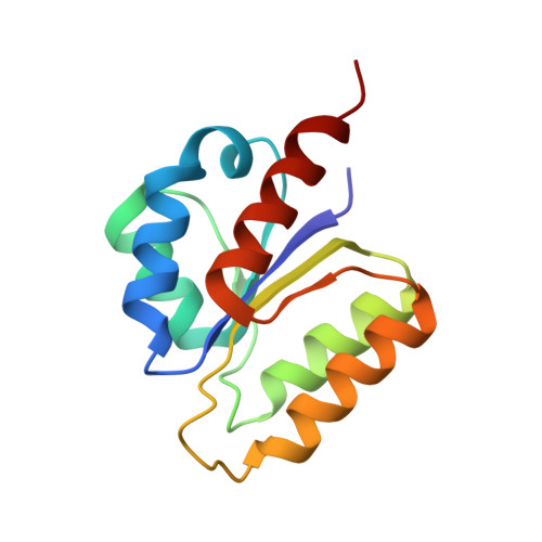 | |
UniProt | |||||
Find proteins for Q5SHD6 (Thermus thermophilus (strain ATCC 27634 / DSM 579 / HB8)) Explore Q5SHD6 Go to UniProtKB: Q5SHD6 | |||||
Entity Groups | |||||
| Sequence Clusters | 30% Identity50% Identity70% Identity90% Identity95% Identity100% Identity | ||||
| UniProt Group | Q5SHD6 | ||||
Sequence AnnotationsExpand | |||||
| |||||
| Ligands 1 Unique | |||||
|---|---|---|---|---|---|
| ID | Chains | Name / Formula / InChI Key | 2D Diagram | 3D Interactions | |
| SO4 Query on SO4 | G [auth A] H [auth B] I [auth C] J [auth D] K [auth D] | SULFATE ION O4 S QAOWNCQODCNURD-UHFFFAOYSA-L |  | ||
| Length ( Å ) | Angle ( ˚ ) |
|---|---|
| a = 130.612 | α = 90 |
| b = 130.612 | β = 90 |
| c = 96.653 | γ = 120 |
| Software Name | Purpose |
|---|---|
| HKL-2000 | data collection |
| SCALEPACK | data scaling |
| SOLVE | phasing |
| CNS | refinement |
| HKL-2000 | data reduction |
RCSB PDB (citation) is hosted by
RCSB PDB is a member of the

