1WM1

Crystal Structure of Prolyl Aminopeptidase, Complex with Pro-TBODA


Experimental Data Snapshot

  • Method: X-RAY DIFFRACTION
  • Resolution: 2.10 Å
  • R-Value Free: 0.230 
  • R-Value Work: 0.200 

wwPDB Validation   3D Report Full Report


Ligand Structure Quality Assessment 


This is version 1.3 of the entry. See complete history


Literature

Novel inhibitor for prolyl aminopeptidase from Serratia marcescens and studies on the mechanism of substrate recognition of the enzyme using the inhibitor

Inoue, T.Ito, K.Tozaka, T.Hatakeyama, S.Tanaka, N.Nakamura, K.T.Yoshimoto, T.

(2003) Arch Biochem Biophys 416: 147-154

  • DOI: https://doi.org/10.1016/s0003-9861(03)00293-5
  • Primary Citation of Related Structures:  
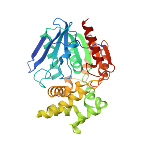
    1WM1

  • PubMed Abstract: 

    Prolyl aminopeptidase from Serratia marcescens hydrolyzed x-beta-naphthylamides (x=prolyl, alanyl, sarcosinyl, L-alpha-aminobutylyl, and norvalyl), which suggested that the enzyme has a pocket for a five-member ring. Based on the substrate specificity, novel inhibitors of Pro, Ala, and Sar having 2-tert-butyl-[1,3,4]oxadiazole (TBODA) were synthesized. The K(i) value of Pro-TBODA, Ala-TBODA, and Sar-TBODA was 0.5 microM, 1.6 microM, and 12mM, respectively. The crystal structure of enzyme-Pro-TBODA complex was determined. Pro-TBODA was located at the active site. Four electrostatic interactions were located between the enzyme and the amino group of Pro inhibitors (Glu204:0E1-N:Inh, Glu204:0E2-N:Inh, Glu232:0E1-N:Inh, and Gly46:O-N:Inh), and the residue of the inhibitors was inserted into the hydrophobic pocket composed of Phe139, Leu141, Leu146, Tyr149, Tyr150, and Phe236. The roles of Phe139, Tyr149, and Phe236 in the hydrophobic pocket and Glu204 and Glu232 in the electrostatic interactions were confirmed by site-directed mutagenesis, which indicated that the molecular recognition of proline is achieved through four electrostatic interactions and an insertion in the hydrophobic pocket of the enzyme.


  • Organizational Affiliation

    Graduate School of Biomedical Sciences, Nagasaki University, 1-14 Bunkyo-machi, Nagasaki 852-8521, Japan.


Macromolecules
Find similar proteins by:  (by identity cutoff)  |  3D Structure
Entity ID: 1
MoleculeChains Sequence LengthOrganismDetailsImage
Proline iminopeptidase317Serratia marcescensMutation(s): 0 
EC: 3.4.11.5
UniProt
Find proteins for O32449 (Serratia marcescens)
Explore O32449 
Go to UniProtKB:  O32449
Entity Groups  
Sequence Clusters30% Identity50% Identity70% Identity90% Identity95% Identity100% Identity
UniProt GroupO32449
Sequence Annotations
Expand
  • Reference Sequence
Small Molecules
Ligands 1 Unique
IDChains Name / Formula / InChI Key2D Diagram3D Interactions
PTB
Query on PTB

Download Ideal Coordinates CCD File 
B [auth A](5-TERT-BUTYL-1,3,4-OXADIAZOL-2-YL)[(2R)-PYRROLIDIN-2-YL]METHANONE
C11 H17 N3 O2
YUKGJYBQODPVBL-ZETCQYMHSA-N
Binding Affinity Annotations 
IDSourceBinding Affinity
PTB Binding MOAD:  1WM1 Ki: 500 (nM) from 1 assay(s)
BindingDB:  1WM1 Ki: 500 (nM) from 1 assay(s)
PDBBind:  1WM1 Ki: 500 (nM) from 1 assay(s)
Experimental Data & Validation

Experimental Data

  • Method: X-RAY DIFFRACTION
  • Resolution: 2.10 Å
  • R-Value Free: 0.230 
  • R-Value Work: 0.200 
  • Space Group: P 43 21 2
Unit Cell:
Length ( Å )Angle ( ˚ )
a = 65.445α = 90
b = 65.445β = 90
c = 169.071γ = 90
Software Package:
Software NamePurpose
MOSFLMdata reduction
SCALAdata scaling
CNSrefinement
CCP4data scaling
CNSphasing

Structure Validation

View Full Validation Report


Ligand Structure Quality Assessment 


Entry History 

Deposition Data

Revision History  (Full details and data files)

  • Version 1.0: 2004-07-20
    Type: Initial release
  • Version 1.1: 2008-04-30
    Changes: Version format compliance
  • Version 1.2: 2011-07-13
    Changes: Version format compliance
  • Version 1.3: 2024-03-13
    Changes: Data collection, Database references, Derived calculations