Determination of the role of the Carboxyl-terminal leucine-122 in FMN-binding protein by mutational and structural analysis.
Kitamura, M., Terakawa, K., Inoue, H., Hayashida, T., Suto, K., Morimoto, Y., Yasuoka, N., Shibata, N., Higuchi, Y.(2007) J Biochem 141: 459-468
- PubMed: 17261542
- DOI: https://doi.org/10.1093/jb/mvm051
- Primary Citation of Related Structures:
1WLI, 1WLK, 3A20 - PubMed Abstract:
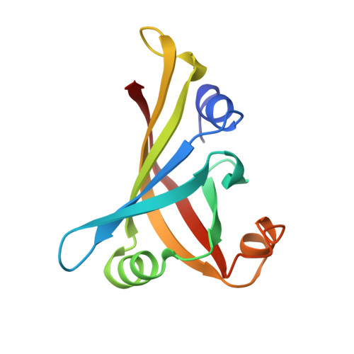
Mutants of flavin mononucleotide-binding protein (FMN-bp) were made by site-directed mutagenesis to investigate the role of carboxyl-terminal Leu122 of the pairing subunit in controlling redox potentials, binding the prosthetic group, and forming the tertiary and quaternary structure. We compared the oxidation-reduction potentials, FMN-binding properties, and higher structures of wild-type FMN-bp and four mutant proteins (L122Y, L122E, L122K and L122-deleted). We found that the redox potentials were affected by mutations. Also, the affinities of L122E, L122K and L122 deletion mutant apoproteins for FMN were lower than for the wild-type apoprotein, whereas the affinity of L122Y for FMN was increased. Analytical ultracentrifugation showed that the dissociation constants for dimerization of L122E and L122K were larger than for wild-type FMN-bp, whereas the dissociation constants for L122Y and the deletion mutant were lower than for the wild type. Finally, we determined the higher structures of L122Y, L122E and L122K mutants by X-ray crystallography. Our results show that the mutation of Leu122 in FMN-bp changes midpoint potentials, dissociation constants for FMN, and dimer formation, indicating that this residue is important in the pairing subunit.
Organizational Affiliation:
Department of Applied and Bioapplied Chemistry, Graduate School of Engineering, Osaka City University, Sugimoto, Osaka, Japan. kitamura@bioa.eng.osaka-cu.ac.jp