Crystal Structure of Bacillus subtilis Guanine Deaminase: THE FIRST DOMAIN-SWAPPED STRUCTURE IN THE CYTIDINE DEAMINASE SUPERFAMILY
Liaw, S.H., Chang, Y.J., Lai, C.T., Chang, H.C., Chang, G.G.(2004) J Biol Chem 279: 35479-35485
- PubMed: 15180998
- DOI: https://doi.org/10.1074/jbc.M405304200
- Primary Citation of Related Structures:
1WKQ - PubMed Abstract:
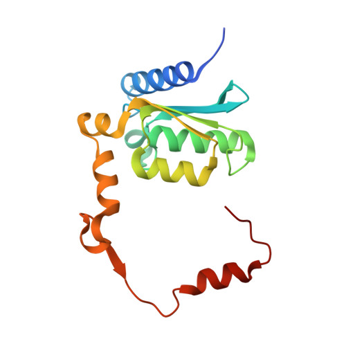
Guanine deaminase, a key enzyme in the nucleotide metabolism, catalyzes the hydrolytic deamination of guanine into xanthine. The crystal structure of the 156-residue guanine deaminase from Bacillus subtilis has been solved at 1.17-A resolution. Unexpectedly, the C-terminal segment is swapped to form an intersubunit active site and an intertwined dimer with an extensive interface of 3900 A(2) per monomer. The essential zinc ion is ligated by a water molecule together with His(53), Cys(83), and Cys(86). A transition state analog was modeled into the active site cavity based on the tightly bound imidazole and water molecules, allowing identification of the conserved deamination mechanism and specific substrate recognition by Asp(114) and Tyr(156'). The closed conformation also reveals that substrate binding seals the active site entrance, which is controlled by the C-terminal tail. Therefore, the domain swapping has not only facilitated the dimerization but has also ensured specific substrate recognition. Finally, a detailed structural comparison of the cytidine deaminase superfamily illustrates the functional versatility of the divergent active sites found in the guanine, cytosine, and cytidine deaminases and suggests putative specific substrate-interacting residues for other members such as dCMP deaminases.
Organizational Affiliation:
Structural Biology Program, Faculty of Life Science, Institute of Biotechnology in Medicine, and Institute of Genetics, National Yang-Ming University, Taipei 11221, Taiwan. shliaw@ym.edu.tw