1WCY

Crystal Structure Of Human Dipeptidyl Peptidase IV (DPPIV) Complex With Diprotin A


Experimental Data Snapshot

  • Method: X-RAY DIFFRACTION
  • Resolution: 2.20 Å
  • R-Value Free: 0.265 
  • R-Value Work: 0.224 

wwPDB Validation   3D Report Full Report


This is version 2.1 of the entry. See complete history


Literature

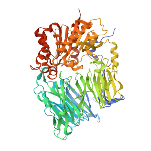
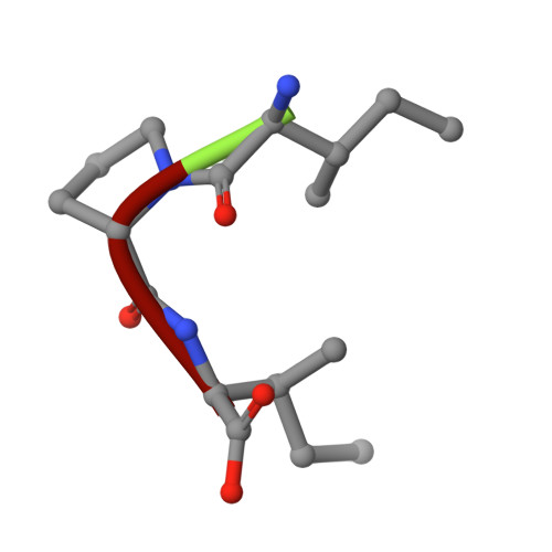
The crystal structure of human dipeptidyl peptidase IV (DPPIV) complex with diprotin A

Hiramatsu, H.Yamamoto, A.Kyono, K.Higashiyama, Y.Fukushima, C.Shima, H.Sugiyama, S.Inaka, K.Shimizu, R.

(2004) Biol Chem 385: 561-564

  • DOI: https://doi.org/10.1515/BC.2004.068
  • Primary Citation of Related Structures:  
    1WCY

  • PubMed Abstract: 

    Dipeptidyl peptidase IV (DPPIV) is a serine protease, a member of the prolyl oligopeptidase (POP) family, and has been implicated in several diseases. Therefore, it seems important to develop selective inhibitors for human DPPIV (hDPPIV) that are able to control the biological function of hDPPIV. In order to elucidate the binding mode and substrate specificity, we determined the crystal structure complex of hDPPIV and diprotin A (IIe-Pro-IIe), a slowly hydrolyzed substrate of hDPPIV, at 2.2 A resolution. In this paper, we discuss the molecular interaction mechanism of diprotin A with hDPPIV based on the X-ray crystal structure.


  • Organizational Affiliation

    Tanabe Seiyaku Co. Ltd., 3-16-89 Kashima, Yodogawa-ku, Osaka 532-8505, Japan.


Macromolecules
Find similar proteins by:  (by identity cutoff)  |  3D Structure
Entity ID: 1
MoleculeChains Sequence LengthOrganismDetailsImage
Dipeptidyl peptidase IV
A, B
740Homo sapiensMutation(s): 0 
EC: 3.4.14.5
UniProt & NIH Common Fund Data Resources
Find proteins for P27487 (Homo sapiens)
Explore P27487 
Go to UniProtKB:  P27487
PHAROS:  P27487
GTEx:  ENSG00000197635 
Entity Groups  
Sequence Clusters30% Identity50% Identity70% Identity90% Identity95% Identity100% Identity
UniProt GroupP27487
Sequence Annotations
Expand
  • Reference Sequence

Find similar proteins by:  Sequence   |   3D Structure  

Entity ID: 2
MoleculeChains Sequence LengthOrganismDetailsImage
Diprotin A
C, D
3N/AMutation(s): 0 
Sequence Annotations
Expand
  • Reference Sequence
Oligosaccharides

Help

Entity ID: 3
MoleculeChains Length2D Diagram Glycosylation3D Interactions
alpha-L-fucopyranose-(1-6)-2-acetamido-2-deoxy-beta-D-glucopyranose
E, I
2N-Glycosylation
Glycosylation Resources
GlyTouCan:  G86851RC
GlyCosmos:  G86851RC
GlyGen:  G86851RC
Entity ID: 4
MoleculeChains Length2D Diagram Glycosylation3D Interactions
2-acetamido-2-deoxy-beta-D-glucopyranose-(1-4)-2-acetamido-2-deoxy-beta-D-glucopyranose
F, G, H, J, K
2N-Glycosylation
Glycosylation Resources
GlyTouCan:  G42666HT
GlyCosmos:  G42666HT
GlyGen:  G42666HT
Experimental Data & Validation

Experimental Data

  • Method: X-RAY DIFFRACTION
  • Resolution: 2.20 Å
  • R-Value Free: 0.265 
  • R-Value Work: 0.224 
  • Space Group: P 21 21 21
Unit Cell:
Length ( Å )Angle ( ˚ )
a = 118.02α = 90
b = 126.06β = 90
c = 136.67γ = 90
Software Package:
Software NamePurpose
MOSFLMdata reduction
SCALAdata scaling
CNXrefinement
CCP4data scaling
CNXphasing

Structure Validation

View Full Validation Report


Entry History 

Deposition Data

Revision History  (Full details and data files)

  • Version 1.0: 2005-05-07
    Type: Initial release
  • Version 1.1: 2008-04-30
    Changes: Version format compliance
  • Version 1.2: 2011-07-13
    Changes: Non-polymer description, Version format compliance
  • Version 2.0: 2020-07-29
    Type: Remediation
    Reason: Carbohydrate remediation
    Changes: Atomic model, Data collection, Database references, Derived calculations, Structure summary
  • Version 2.1: 2023-10-25
    Changes: Data collection, Database references, Refinement description, Structure summary