1WCF

1.54 A CRYSTAL STRUCTURE OF RV3628, MYCOBACTERIUM TUBERCULOSIS INORGANIC PYROPHOSPHATASE (PPASE) AT PH7.0


Experimental Data Snapshot

  • Method: X-RAY DIFFRACTION
  • Resolution: 1.54 Å
  • R-Value Free: 0.170 
  • R-Value Work: 0.156 
  • R-Value Observed: 0.157 

wwPDB Validation   3D Report Full Report


This is version 2.0 of the entry. See complete history


Literature

Structure of the Mycobacterium Tuberculosis Soluble Inorganic Pyrophosphatase Rv3628 at Ph 7.0.

Benini, S.Wilson, K.S.

(2011) Acta Crystallogr Sect F Struct Biol Cryst Commun 67: 866

  • DOI: https://doi.org/10.1107/S1744309111023323
  • Primary Citation of Related Structures:  
    1WCF

  • PubMed Abstract: 

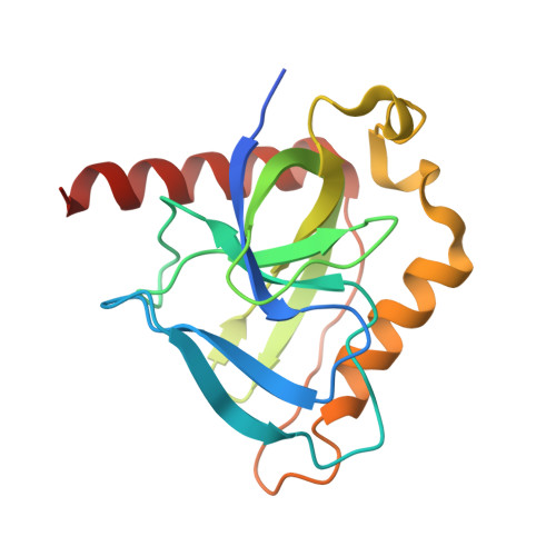
    The 1.5 Å resolution crystal structure of the Mycobacterium tuberculosis soluble inorganic pyrophosphatase Rv3628 at pH 7.0 is reported. The M. tuberculosis and M. leprae genomes include genes for the only two family I inorganic pyrophosphatases known to contain two histidines in the active site. The role of these two residues in catalysis is not fully understood. Mutational and functional studies of the M. tuberculosis enzyme showed that His21 and His86 are not essential for pyrophosphate hydrolysis, but are responsible for a shift in the optimal pH for the reaction compared with the Escherichia coli enzyme. Comparison with the structure previously reported at pH 5.0 provides further insight into the role of the two histidines. Two potassium-binding sites are found as a result of the high potassium concentration in the mother liquor.


  • Organizational Affiliation

    Faculty of Science and Technology, Free University of Bolzano, Bolzano, Italy. stefano.benini@unibz.it


Macromolecules
Find similar proteins by:  (by identity cutoff)  |  3D Structure
Entity ID: 1
MoleculeChains Sequence LengthOrganismDetailsImage
INORGANIC PYROPHOSPHATASE171Mycobacterium tuberculosis H37RvMutation(s): 0 
EC: 3.6.1.1
UniProt
Find proteins for P9WI55 (Mycobacterium tuberculosis (strain ATCC 25618 / H37Rv))
Explore P9WI55 
Go to UniProtKB:  P9WI55
Entity Groups  
Sequence Clusters30% Identity50% Identity70% Identity90% Identity95% Identity100% Identity
UniProt GroupP9WI55
Sequence Annotations
Expand
  • Reference Sequence
Experimental Data & Validation

Experimental Data

  • Method: X-RAY DIFFRACTION
  • Resolution: 1.54 Å
  • R-Value Free: 0.170 
  • R-Value Work: 0.156 
  • R-Value Observed: 0.157 
  • Space Group: P 63 2 2
Unit Cell:
Length ( Å )Angle ( ˚ )
a = 96.902α = 90
b = 96.902β = 90
c = 103.312γ = 120
Software Package:
Software NamePurpose
REFMACrefinement
DENZOdata reduction
SCALEPACKdata scaling
AMoREphasing

Structure Validation

View Full Validation Report


Entry History 

Deposition Data

Revision History  (Full details and data files)

  • Version 1.0: 2006-07-12
    Type: Initial release
  • Version 1.1: 2011-08-17
    Changes: Atomic model, Database references, Derived calculations, Non-polymer description, Other, Structure summary, Version format compliance
  • Version 2.0: 2023-12-13
    Changes: Atomic model, Data collection, Database references, Derived calculations, Other, Refinement description