1WAV

CRYSTAL STRUCTURE OF FORM B MONOCLINIC CRYSTAL OF INSULIN


Experimental Data Snapshot

  • Method: X-RAY DIFFRACTION
  • Resolution: 2.50 Å
  • R-Value Work: 0.206 
  • R-Value Observed: 0.206 

wwPDB Validation   3D Report Full Report


This is version 1.2 of the entry. See complete history


Literature

Molecular replacement study on form-B monoclinic crystal of insulin.

Ding, J.Wan, Z.Chang, W.Liang, D.

(1996) Sci China C Life Sci 39: 144-153

  • Primary Citation of Related Structures:  
    1WAV

  • PubMed Abstract: 

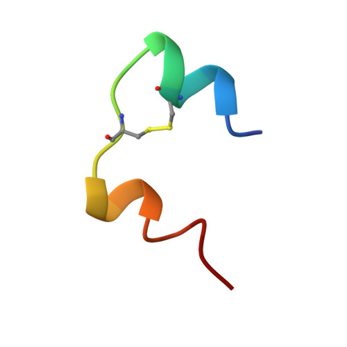
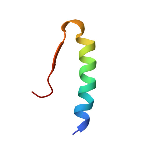
    The form-B monoclinic insulin crystal was obtained from the sodium citrate buffer with 1% zinc chloride, keeping phenolic content between 0.76% and 1.25%. Its space group is P2(1), cell constants are: a = 4.924 nm, b = 6.094 nm, c = 4.818 nm, beta = 95.8 degrees. There are 6 insulin molecules which form a hexamer. The initial phase was obtained by using rotation function program of X-PLOR program package and molecular packing program of our laboratory. The molecular model was chosen from 4 zinc bovine insulin hexamer. After the preliminary refinement by using the macromolecular rigid body refinement technique, the molecular model was further refined and adjusted by using the energy-minimizing stereochemically restrained least-squared refinement on the difference Fourier maps. The final R-factor is 22.4% at 0.3 nm resolution, the r.m.s. deviations from standard bond length and bond angle are 0.0022 nm and 4.7 degrees, respectively.


  • Organizational Affiliation

    State Key Laboratory of Biomacromolecules, Institute of Biophysics, Chinese Academy of Sciences, Beijing, China.


Macromolecules

Find similar proteins by:  Sequence   |   3D Structure  

Entity ID: 1
MoleculeChains Sequence LengthOrganismDetailsImage
INSULIN
A, C, E, G, I
A, C, E, G, I, K
21Sus scrofaMutation(s): 0 
UniProt
Find proteins for P01315 (Sus scrofa)
Explore P01315 
Go to UniProtKB:  P01315
Entity Groups  
Sequence Clusters30% Identity50% Identity70% Identity90% Identity95% Identity100% Identity
UniProt GroupP01315
Sequence Annotations
Expand
  • Reference Sequence
Find similar proteins by:  (by identity cutoff)  |  3D Structure
Entity ID: 2
MoleculeChains Sequence LengthOrganismDetailsImage
INSULIN
B, D, F, H, J
B, D, F, H, J, L
30Sus scrofaMutation(s): 0 
UniProt
Find proteins for P01315 (Sus scrofa)
Explore P01315 
Go to UniProtKB:  P01315
Entity Groups  
Sequence Clusters30% Identity50% Identity70% Identity90% Identity95% Identity100% Identity
UniProt GroupP01315
Sequence Annotations
Expand
  • Reference Sequence
Experimental Data & Validation

Experimental Data

  • Method: X-RAY DIFFRACTION
  • Resolution: 2.50 Å
  • R-Value Work: 0.206 
  • R-Value Observed: 0.206 
  • Space Group: P 1 21 1
Unit Cell:
Length ( Å )Angle ( ˚ )
a = 49.24α = 90
b = 60.94β = 95.8
c = 48.18γ = 90
Software Package:
Software NamePurpose
WEISdata collection
X-PLORmodel building
X-PLORrefinement
WEISdata reduction
X-PLORphasing

Structure Validation

View Full Validation Report


Entry History 

Deposition Data

Revision History  (Full details and data files)

  • Version 1.0: 1997-02-28
    Type: Initial release
  • Version 1.1: 2008-03-03
    Changes: Version format compliance
  • Version 1.2: 2011-07-13
    Changes: Version format compliance