1WAJ

DNA POLYMERASE FROM BACTERIOPHAGE RB69


Experimental Data Snapshot

  • Method: X-RAY DIFFRACTION
  • Resolution: 2.80 Å
  • R-Value Free: 0.280 
  • R-Value Work: 0.210 
  • R-Value Observed: 0.210 

wwPDB Validation   3D Report Full Report


Ligand Structure Quality Assessment 


This is version 1.3 of the entry. See complete history


Literature

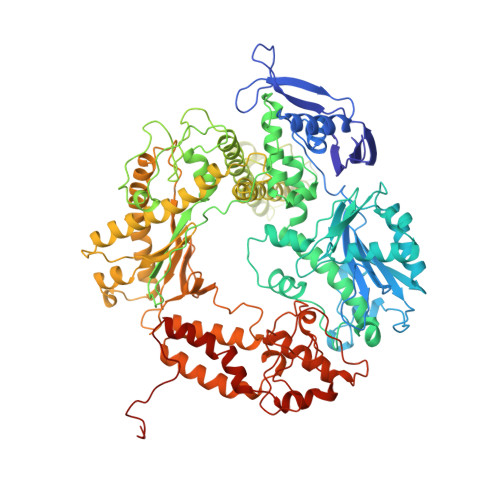
Crystal structure of a pol alpha family replication DNA polymerase from bacteriophage RB69.

Wang, J.Sattar, A.K.Wang, C.C.Karam, J.D.Konigsberg, W.H.Steitz, T.A.

(1997) Cell 89: 1087-1099

  • DOI: https://doi.org/10.1016/s0092-8674(00)80296-2
  • Primary Citation of Related Structures:  
    1WAF, 1WAJ

  • PubMed Abstract: 

    The 2.8 A resolution crystal structure of the bacteriophage RB69 gp43, a member of the eukaryotic pol alpha family of replicative DNA polymerases, shares some similarities with other polymerases but shows many differences. Although its palm domain has the same topology as other polymerases, except rat DNA polymerase beta, one of the three carboxylates required for nucleotidyl transfer is located on a different beta strand. The structures of the fingers and thumb domains are unrelated to all other known polymerase structures. The editing 3'-5' exonuclease domain of gp43 is homologous to that of E. coli DNA polymerase I but lies on the opposite side of the polymerase active site. An extended structure-based alignment of eukaryotic DNA polymerase sequences provides structural insights that should be applicable to most eukaryotic DNA polymerases.


  • Organizational Affiliation

    Department of Molecular Biophysics and Biochemistry, Yale University, New Haven, Connecticut 06520-8114, USA.


Macromolecules
Find similar proteins by:  (by identity cutoff)  |  3D Structure
Entity ID: 1
MoleculeChains Sequence LengthOrganismDetailsImage
DNA POLYMERASE903Escherichia phage RB69Mutation(s): 0 
Gene Names: POL
EC: 2.7.7.7
UniProt
Find proteins for Q38087 (Escherichia phage RB69)
Explore Q38087 
Go to UniProtKB:  Q38087
Entity Groups  
Sequence Clusters30% Identity50% Identity70% Identity90% Identity95% Identity100% Identity
UniProt GroupQ38087
Sequence Annotations
Expand
  • Reference Sequence
Small Molecules
Ligands 1 Unique
IDChains Name / Formula / InChI Key2D Diagram3D Interactions
5GP
Query on 5GP

Download Ideal Coordinates CCD File 
B [auth A]GUANOSINE-5'-MONOPHOSPHATE
C10 H14 N5 O8 P
RQFCJASXJCIDSX-UUOKFMHZSA-N
Experimental Data & Validation

Experimental Data

  • Method: X-RAY DIFFRACTION
  • Resolution: 2.80 Å
  • R-Value Free: 0.280 
  • R-Value Work: 0.210 
  • R-Value Observed: 0.210 
  • Space Group: P 21 21 21
Unit Cell:
Length ( Å )Angle ( ˚ )
a = 81.41α = 90
b = 117.6β = 90
c = 199.24γ = 90
Software Package:
Software NamePurpose
X-PLORmodel building
X-PLORrefinement
DENZOdata reduction
SCALEPACKdata scaling
X-PLORphasing

Structure Validation

View Full Validation Report


Ligand Structure Quality Assessment 


Entry History 

Deposition Data

Revision History  (Full details and data files)

  • Version 1.0: 1998-01-14
    Type: Initial release
  • Version 1.1: 2008-03-24
    Changes: Version format compliance
  • Version 1.2: 2011-07-13
    Changes: Version format compliance
  • Version 1.3: 2024-02-14
    Changes: Data collection, Database references, Derived calculations, Other