1WA9

Crystal Structure of the PAS repeat region of the Drosophila clock protein PERIOD


Experimental Data Snapshot

  • Method: X-RAY DIFFRACTION
  • Resolution: 3.15 Å
  • R-Value Free: 0.277 
  • R-Value Work: 0.228 
  • R-Value Observed: 0.228 

wwPDB Validation   3D Report Full Report


This is version 1.2 of the entry. See complete history


Literature

Crystal Structure and Interactions of the Pas Repeat Region of the Drosophila Clock Protein Period

Yildiz, O.Doi, M.Yujnovsky, I.Cardone, L.Berndt, A.Hennig, S.Schulze, S.Urbanke, C.Sassone-Corsi, P.Wolf, E.

(2005) Mol Cell 17: 69

  • DOI: https://doi.org/10.1016/j.molcel.2004.11.022
  • Primary Citation of Related Structures:  
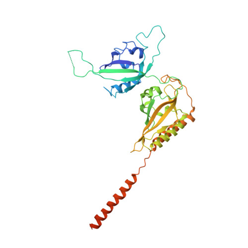
    1WA9

  • PubMed Abstract: 

    PERIOD proteins are central components of the Drosophila and mammalian circadian clock. Their function is controlled by daily changes in synthesis, cellular localization, phosphorylation, degradation, as well as specific interactions with other clock components. Here we present the crystal structure of a Drosophila PERIOD (dPER) fragment comprising two tandemly organized PAS (PER-ARNT-SIM) domains (PAS-A and PAS-B) and two additional C-terminal alpha helices (alphaE and alphaF). Our analysis reveals a noncrystallographic dPER dimer mediated by intermolecular interactions of PAS-A with PAS-B and helix alphaF. We show that alphaF is essential for dPER homodimerization and that the PAS-A-alphaF interaction plays a crucial role in dPER clock function, as it is affected by the 29 hr long-period perL mutation.


  • Organizational Affiliation

    Department of Structural Biology, Max-Planck-Institute for Molecular Physiology, Otto-Hahn-Strasse 11, 44227 Dortmund, Germany.


Macromolecules
Find similar proteins by:  (by identity cutoff)  |  3D Structure
Entity ID: 1
MoleculeChains Sequence LengthOrganismDetailsImage
PERIOD CIRCADIAN PROTEIN
A, B
368Drosophila melanogasterMutation(s): 0 
UniProt
Find proteins for P07663 (Drosophila melanogaster)
Explore P07663 
Go to UniProtKB:  P07663
Entity Groups  
Sequence Clusters30% Identity50% Identity70% Identity90% Identity95% Identity100% Identity
UniProt GroupP07663
Sequence Annotations
Expand
  • Reference Sequence
Experimental Data & Validation

Experimental Data

  • Method: X-RAY DIFFRACTION
  • Resolution: 3.15 Å
  • R-Value Free: 0.277 
  • R-Value Work: 0.228 
  • R-Value Observed: 0.228 
  • Space Group: P 43
Unit Cell:
Length ( Å )Angle ( ˚ )
a = 140.44α = 90
b = 140.44β = 90
c = 61.45γ = 90
Software Package:
Software NamePurpose
CNSrefinement
XDSdata reduction
XSCALEdata scaling
CNSphasing

Structure Validation

View Full Validation Report


Entry History 

Deposition Data

Revision History  (Full details and data files)

  • Version 1.0: 2005-01-12
    Type: Initial release
  • Version 1.1: 2011-05-08
    Changes: Version format compliance
  • Version 1.2: 2011-07-13
    Changes: Version format compliance