1W8Y

Crystal structure of the nitrocefin acyl-DD-peptidase from Actinomadura R39.


Experimental Data Snapshot

  • Method: X-RAY DIFFRACTION
  • Resolution: 2.40 Å
  • R-Value Free: 0.277 
  • R-Value Work: 0.220 
  • R-Value Observed: 0.220 

wwPDB Validation   3D Report Full Report


Ligand Structure Quality Assessment 


This is version 2.1 of the entry. See complete history


Literature

Crystal Structure of the Actinomadura R39 Dd- Peptidase Reveals New Domains in Penicillin- Binding Proteins.

Sauvage, E.Herman, R.Petrella, S.Duez, C.Bouillenne, F.Frere, J.M.Charlier, P.

(2005) J Biol Chem 280: 31249

  • DOI: https://doi.org/10.1074/jbc.M503271200
  • Primary Citation of Related Structures:  
    1W79, 1W8Q, 1W8Y

  • PubMed Abstract: 

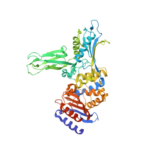
    Actinomadura sp. R39 produces an exocellular DD-peptidase/penicillin-binding protein (PBP) whose primary structure is similar to that of Escherichia coli PBP4. It is characterized by a high beta-lactam-binding activity (second order rate constant for the acylation of the active site serine by benzylpenicillin: k2/K = 300 mm(-1) s(-1)). The crystal structure of the DD-peptidase from Actinomadura R39 was solved at a resolution of 1.8 angstroms by single anomalous dispersion at the cobalt resonance wavelength. The structure is composed of three domains: a penicillin-binding domain similar to the penicillin-binding domain of E. coli PBP5 and two domains of unknown function. In most multimodular PBPs, additional domains are generally located at the C or N termini of the penicillin-binding domain. In R39, the other two domains are inserted in the penicillin-binding domain, between the SXXK and SXN motifs, in a manner similar to "Matryoshka dolls." One of these domains is composed of a five-stranded beta-sheet with two helices on one side, and the other domain is a double three-stranded beta-sheet inserted in the previous domain. Additionally, the 2.4-angstroms structure of the acyl-enzyme complex of R39 with nitrocefin reveals the absence of active site conformational change upon binding the beta-lactams.


  • Organizational Affiliation

    Centre d'Ingénierie des Protéines, Université de Liège, Institut de Physique B5, B-4000 Liège, Belgium. eric.sauvage@ulg.ac.be


Macromolecules
Find similar proteins by:  (by identity cutoff)  |  3D Structure
Entity ID: 1
MoleculeChains Sequence LengthOrganismDetailsImage
D-alanyl-D-alanine carboxypeptidase
A, B, C, D
489Actinomadura sp. R39Mutation(s): 0 
EC: 3.4.16.4
UniProt
Find proteins for P39045 (Actinomadura sp. (strain R39))
Explore P39045 
Go to UniProtKB:  P39045
Entity Groups  
Sequence Clusters30% Identity50% Identity70% Identity90% Identity95% Identity100% Identity
UniProt GroupP39045
Sequence Annotations
Expand
  • Reference Sequence
Small Molecules
Ligands 3 Unique
IDChains Name / Formula / InChI Key2D Diagram3D Interactions
NCF
Query on NCF

Download Ideal Coordinates CCD File 
BA [auth D],
K [auth A],
P [auth B],
U [auth C]
(2R)-2-{(1R)-2-OXO-1-[(2-THIENYLACETYL)AMINO]ETHYL}-5,6-DIHYDRO-2H-1,3-THIAZINE-4-CARBOXYLIC ACID
C13 H14 N2 O5 S2
LNPNJPUNJYVKHH-WDEREUQCSA-N
SO4
Query on SO4

Download Ideal Coordinates CCD File 
E [auth A]
F [auth A]
G [auth A]
H [auth A]
L [auth B]
E [auth A],
F [auth A],
G [auth A],
H [auth A],
L [auth B],
M [auth B],
N [auth B],
O [auth B],
Q [auth C],
R [auth C],
S [auth C],
T [auth C],
V [auth D],
W [auth D],
X [auth D],
Y [auth D]
SULFATE ION
O4 S
QAOWNCQODCNURD-UHFFFAOYSA-L
MG
Query on MG

Download Ideal Coordinates CCD File 
AA [auth D],
I [auth A],
J [auth A],
Z [auth D]
MAGNESIUM ION
Mg
JLVVSXFLKOJNIY-UHFFFAOYSA-N
Experimental Data & Validation

Experimental Data

  • Method: X-RAY DIFFRACTION
  • Resolution: 2.40 Å
  • R-Value Free: 0.277 
  • R-Value Work: 0.220 
  • R-Value Observed: 0.220 
  • Space Group: P 1 21 1
Unit Cell:
Length ( Å )Angle ( ˚ )
a = 103.49α = 90
b = 94.36β = 94.58
c = 107.2γ = 90
Software Package:
Software NamePurpose
CNSrefinement
MOSFLMdata reduction
SCALAdata scaling

Structure Validation

View Full Validation Report


Ligand Structure Quality Assessment 


Entry History 

Deposition Data

Revision History  (Full details and data files)

  • Version 1.0: 2005-06-28
    Type: Initial release
  • Version 1.1: 2011-07-13
    Changes: Source and taxonomy, Version format compliance
  • Version 2.0: 2021-09-15
    Changes: Atomic model, Database references, Derived calculations, Non-polymer description, Other, Source and taxonomy, Structure summary
  • Version 2.1: 2023-12-13
    Changes: Data collection, Refinement description