1W8K

Crystal structure of apical membrane antigen 1 from Plasmodium vivax


Experimental Data Snapshot

  • Method: X-RAY DIFFRACTION
  • Resolution: 1.80 Å
  • R-Value Free: 0.269 
  • R-Value Work: 0.219 
  • R-Value Observed: 0.222 

wwPDB Validation   3D Report Full Report


This is version 1.2 of the entry. See complete history


Literature

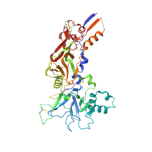
Crystal Structure of the Malaria Vaccine Candidate Apical Membrane Antigen 1

Pizarro, J.C.Vulliez-Le Normand, B.Chesne-Seck, M.-L.Collins, C.Withers-Martinez, C.Hackett, F.Blackman, M.Faber, B.Remarque, E.Kocken, C.H.M.Thomas, A.W.Bentley, G.A.

(2005) Science 308: 408

  • DOI: https://doi.org/10.1126/science.1107449
  • Primary Citation of Related Structures:  
    1W81, 1W8K

  • PubMed Abstract: 

    Apical membrane antigen 1 from Plasmodium is a leading malaria vaccine candidate. The protein is essential for host-cell invasion, but its molecular function is unknown. The crystal structure of the three domains comprising the ectoplasmic region of the antigen from P. vivax, solved at 1.8 angstrom resolution, shows that domains I and II belong to the PAN motif, which defines a superfamily of protein folds implicated in receptor binding. We also mapped the epitope of an invasion-inhibitory monoclonal antibody specific for the P. falciparum ortholog and modeled this to the structure. The location of the epitope and current knowledge on structure-function correlations for PAN domains together suggest a receptor-binding role during invasion in which domain II plays a critical part. These results are likely to aid vaccine and drug design.


  • Organizational Affiliation

    Unité d'Immunologie Structurale, Centre National de la Recherche Scientifique, URA 2185, Institut Pasteur, 25 rue du Docteur Roux, 75724 Paris, France.


Macromolecules
Find similar proteins by:  (by identity cutoff)  |  3D Structure
Entity ID: 1
MoleculeChains Sequence LengthOrganismDetailsImage
APICAL MEMBRANE ANTIGEN 1447Plasmodium vivaxMutation(s): 3 
UniProt
Find proteins for O61130 (Plasmodium vivax)
Explore O61130 
Go to UniProtKB:  O61130
Entity Groups  
Sequence Clusters30% Identity50% Identity70% Identity90% Identity95% Identity100% Identity
UniProt GroupO61130
Sequence Annotations
Expand
  • Reference Sequence
Small Molecules
Ligands 1 Unique
IDChains Name / Formula / InChI Key2D Diagram3D Interactions
IMD
Query on IMD

Download Ideal Coordinates CCD File 
B [auth A]IMIDAZOLE
C3 H5 N2
RAXXELZNTBOGNW-UHFFFAOYSA-O
Experimental Data & Validation

Experimental Data

  • Method: X-RAY DIFFRACTION
  • Resolution: 1.80 Å
  • R-Value Free: 0.269 
  • R-Value Work: 0.219 
  • R-Value Observed: 0.222 
  • Space Group: C 1 2 1
Unit Cell:
Length ( Å )Angle ( ˚ )
a = 150.03α = 90
b = 53.76β = 113.18
c = 60.29γ = 90
Software Package:
Software NamePurpose
REFMACrefinement
XDSdata reduction
XSCALEdata scaling
SHARPphasing

Structure Validation

View Full Validation Report


Entry History 

Deposition Data

Revision History  (Full details and data files)

  • Version 1.0: 2005-03-01
    Type: Initial release
  • Version 1.1: 2011-05-08
    Changes: Version format compliance
  • Version 1.2: 2011-07-13
    Changes: Version format compliance