1W80

Crystal structure of the alpha-adaptin appendage domain, from the AP2 adaptor complex, bound to 2 peptides from Synaptojanin170


Experimental Data Snapshot

  • Method: X-RAY DIFFRACTION
  • Resolution: 1.90 Å
  • R-Value Free: 0.224 
  • R-Value Work: 0.177 
  • R-Value Observed: 0.179 

wwPDB Validation   3D Report Full Report


Ligand Structure Quality Assessment 


This is version 1.3 of the entry. See complete history


Literature

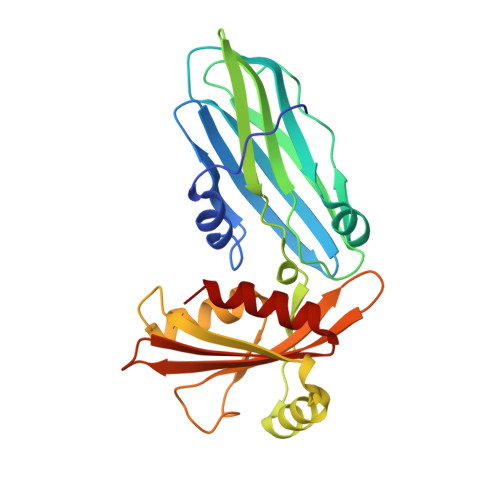
Evolving Nature of the Ap2 Alpha-Appendage Hub During Clathrin-Coated Vesicle Endocytosis.

Praefcke, G.J.K.Ford, M.G.J.Schmid, E.M.Olesen, L.E.Gallop, J.L.Peak-Chew, S.-Y.Vallis, Y.Babu, M.M.Mills, I.G.Mcmahon, H.T.

(2004) EMBO J 23: 4371

  • DOI: https://doi.org/10.1038/sj.emboj.7600445
  • Primary Citation of Related Structures:  
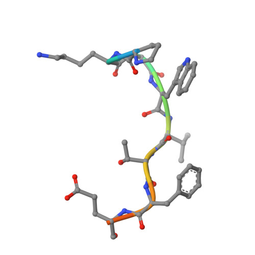
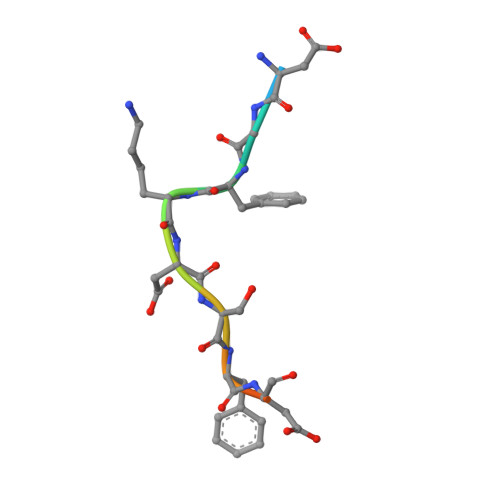
    1W80

  • PubMed Abstract: 

    Clathrin-mediated endocytosis involves the assembly of a network of proteins that select cargo, modify membrane shape and drive invagination, vesicle scission and uncoating. This network is initially assembled around adaptor protein (AP) appendage domains, which are protein interaction hubs. Using crystallography, we show that FxDxF and WVxF peptide motifs from synaptojanin bind to distinct subdomains on alpha-appendages, called 'top' and 'side' sites. Appendages use both these sites to interact with their binding partners in vitro and in vivo. Occupation of both sites simultaneously results in high-affinity reversible interactions with lone appendages (e.g. eps15 and epsin1). Proteins with multiple copies of only one type of motif bind multiple appendages and so will aid adaptor clustering. These clustered alpha(appendage)-hubs have altered properties where they can sample many different binding partners, which in turn can interact with each other and indirectly with clathrin. In the final coated vesicle, most appendage binding partners are absent and thus the functional status of the appendage domain as an interaction hub is temporal and transitory giving directionality to vesicle assembly.


  • Organizational Affiliation

    Medical Research Council Laboratory of Molecular Biology, Cambridge, UK.


Macromolecules
Find similar proteins by:  (by identity cutoff)  |  3D Structure
Entity ID: 1
MoleculeChains Sequence LengthOrganismDetailsImage
ADAPTER-RELATED PROTEIN COMPLEX 2 ALPHA 2 SUBUNIT250Mus musculusMutation(s): 0 
UniProt & NIH Common Fund Data Resources
Find proteins for P17427 (Mus musculus)
Explore P17427 
Go to UniProtKB:  P17427
IMPC:  MGI:101920
Entity Groups  
Sequence Clusters30% Identity50% Identity70% Identity90% Identity95% Identity100% Identity
UniProt GroupP17427
Sequence Annotations
Expand
  • Reference Sequence

Find similar proteins by:  Sequence   |   3D Structure  

Entity ID: 2
MoleculeChains Sequence LengthOrganismDetailsImage
SYNAPTOJANIN 1B [auth P]12Homo sapiensMutation(s): 0 
EC: 3.1.3.36
UniProt & NIH Common Fund Data Resources
Find proteins for O43426 (Homo sapiens)
Explore O43426 
Go to UniProtKB:  O43426
PHAROS:  O43426
GTEx:  ENSG00000159082 
Entity Groups  
Sequence Clusters30% Identity50% Identity70% Identity90% Identity95% Identity100% Identity
UniProt GroupO43426
Sequence Annotations
Expand
  • Reference Sequence

Find similar proteins by:  Sequence   |   3D Structure  

Entity ID: 3
MoleculeChains Sequence LengthOrganismDetailsImage
SYNAPTOJANIN 1C [auth Q]12Homo sapiensMutation(s): 0 
EC: 3.1.3.36
UniProt & NIH Common Fund Data Resources
Find proteins for O43426 (Homo sapiens)
Explore O43426 
Go to UniProtKB:  O43426
PHAROS:  O43426
GTEx:  ENSG00000159082 
Entity Groups  
Sequence Clusters30% Identity50% Identity70% Identity90% Identity95% Identity100% Identity
UniProt GroupO43426
Sequence Annotations
Expand
  • Reference Sequence
Experimental Data & Validation

Experimental Data

  • Method: X-RAY DIFFRACTION
  • Resolution: 1.90 Å
  • R-Value Free: 0.224 
  • R-Value Work: 0.177 
  • R-Value Observed: 0.179 
  • Space Group: C 1 2 1
Unit Cell:
Length ( Å )Angle ( ˚ )
a = 146.599α = 90
b = 67.322β = 94.53
c = 39.721γ = 90
Software Package:
Software NamePurpose
REFMACrefinement
MOSFLMdata reduction
SCALAdata scaling
AMoREphasing

Structure Validation

View Full Validation Report


Ligand Structure Quality Assessment 


Entry History 

Deposition Data

Revision History  (Full details and data files)

  • Version 1.0: 2004-10-27
    Type: Initial release
  • Version 1.1: 2011-05-08
    Changes: Version format compliance
  • Version 1.2: 2011-07-13
    Changes: Version format compliance
  • Version 1.3: 2023-12-13
    Changes: Data collection, Database references, Other, Refinement description