1W7W

Structure and mutational analysis of a plant mitochondrial nucleoside diphosphate kinase: identification of residues involved in serine phosphorylation and oligomerization.


Experimental Data Snapshot

  • Method: X-RAY DIFFRACTION
  • Resolution: 2.80 Å
  • R-Value Free: 0.264 
  • R-Value Work: 0.237 
  • R-Value Observed: 0.237 

wwPDB Validation   3D Report Full Report


This is version 1.4 of the entry. See complete history


Literature

Structure and Mutational Analysis of a Plant Mitochondrial Nucleoside Diphosphate Kinase: Identification of Residues Involved in Serine Phosphorylation and Oligomerisation

Johansson, M.Mackenzie-Hose, A.Andersson, I.Knorpp, C.

(2004) Plant Physiol 136: 3034

  • DOI: https://doi.org/10.1104/pp.104.044040
  • Primary Citation of Related Structures:  
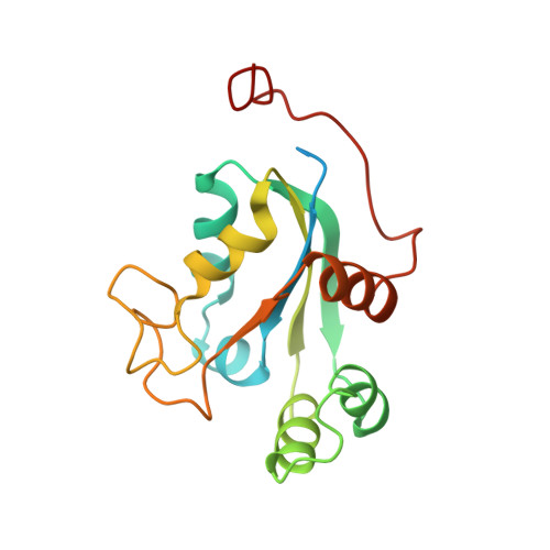
    1W7W

  • PubMed Abstract: 

    We report the first crystal structure of a plant (Pisum sativum L. cv Oregon sugarpod) mitochondrial nucleoside diphosphate kinase. Similar to other eukaryotic nucleoside diphosphate kinases, the plant enzyme is a hexamer; the six monomers in the asymmetric unit are arranged as trimers of dimers. Different functions of the kinase have been correlated with the oligomeric structure and the phosphorylation of Ser residues. We show that the occurrence of Ser autophosphorylation depends on enzymatic activity. The mutation of the strictly conserved Ser-119 to Ala reduced the Ser phosphorylation to about one-half of that observed in wild type with only a modest change of enzyme activity. We also show that mutating another strictly conserved Ser, Ser-69, to Ala reduces the enzyme activity to 6% and 14% of wild-type using dCDP and dTDP as acceptors, respectively. Changes in the oligomerization pattern of the S69A mutant were observed by cross-linking experiments. A reduction in trimer formation and a change in the dimer interaction could be detected with a concomitant increase of tetramers. We conclude that the S69 mutant is involved in the stabilization of the oligomeric state of this plant nucleoside diphosphate kinase.


  • Organizational Affiliation

    Department of Plant Biology and Forest Genetics, Swedish University of Agricultural Sciences, S-750 07 Uppsala, Sweden.


Macromolecules
Find similar proteins by:  (by identity cutoff)  |  3D Structure
Entity ID: 1
MoleculeChains Sequence LengthOrganismDetailsImage
NUCLEOSIDE DIPHOSPHATE KINASE
A, B, C, D, E
A, B, C, D, E, F
182Pisum sativumMutation(s): 0 
EC: 2.7.4.6
UniProt
Find proteins for Q9SP13 (Pisum sativum)
Explore Q9SP13 
Go to UniProtKB:  Q9SP13
Entity Groups  
Sequence Clusters30% Identity50% Identity70% Identity90% Identity95% Identity100% Identity
UniProt GroupQ9SP13
Sequence Annotations
Expand
  • Reference Sequence
Experimental Data & Validation

Experimental Data

  • Method: X-RAY DIFFRACTION
  • Resolution: 2.80 Å
  • R-Value Free: 0.264 
  • R-Value Work: 0.237 
  • R-Value Observed: 0.237 
  • Space Group: P 21 21 21
Unit Cell:
Length ( Å )Angle ( ˚ )
a = 74.1α = 90
b = 84.7β = 90
c = 161.6γ = 90
Software Package:
Software NamePurpose
CNSrefinement
DENZOdata reduction
SCALEPACKdata scaling
MOLREPphasing

Structure Validation

View Full Validation Report


Entry History 

Deposition Data

Revision History  (Full details and data files)

  • Version 1.0: 2004-10-22
    Type: Initial release
  • Version 1.1: 2011-05-08
    Changes: Version format compliance
  • Version 1.2: 2011-07-13
    Changes: Version format compliance
  • Version 1.3: 2017-11-08
    Changes: Source and taxonomy
  • Version 1.4: 2023-12-13
    Changes: Data collection, Database references, Other, Refinement description