1W63

AP1 clathrin adaptor core


Experimental Data Snapshot

  • Method: X-RAY DIFFRACTION
  • Resolution: 4.00 Å
  • R-Value Free: 0.310 
  • R-Value Work: 0.297 
  • R-Value Observed: 0.297 

wwPDB Validation   3D Report Full Report


This is version 1.4 of the entry. See complete history


Literature

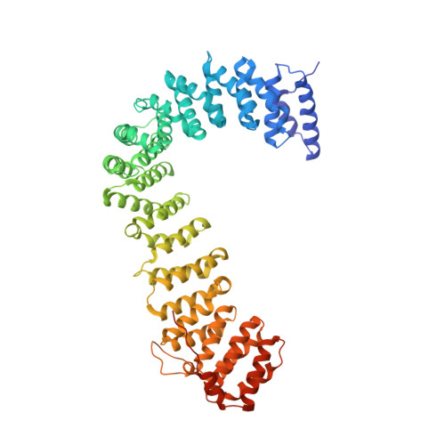
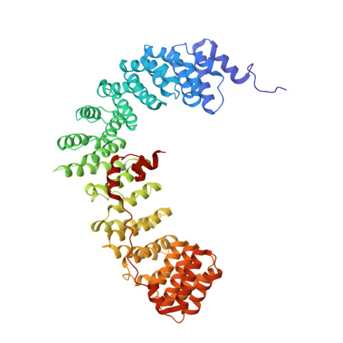
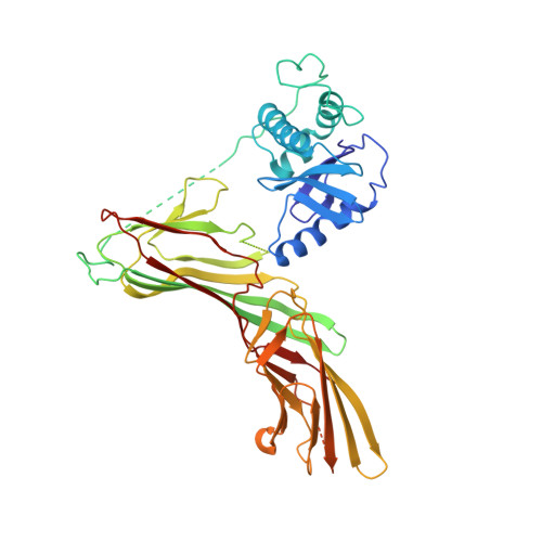
Crystal Structure of the Clathrin Adaptor Protein 1 Core

Heldwein, E.Macia, E.Wang, J.Yin, H.L.Kirchhausen, T.Harrison, S.C.

(2004) Proc Natl Acad Sci U S A 101: 14108

  • DOI: https://doi.org/10.1073/pnas.0406102101
  • Primary Citation of Related Structures:  
    1W63

  • PubMed Abstract: 

    The heterotetrameric adaptor proteins (AP complexes) link the outer lattice of clathrin-coated vesicles with membrane-anchored cargo molecules. We report the crystal structure of the core of the AP-1 complex, which functions in the trans-Golgi network (TGN). Packing of complexes in the crystal generates an exceptionally long (1,135-A) unit-cell axis, but the 6-fold noncrystallographic redundancy yields an excellent map at 4-A resolution. The AP-1 core comprises N-terminal fragments of the two large chains, beta1 and gamma, and the intact medium and small chains, micro1 and sigma1. Its molecular architecture closely resembles that of the core of AP-2, the plasma-membrane-specific adaptor, for which a structure has been determined. Both structures represent an "inactive" conformation with respect to binding of cargo with a tyrosine-based sorting signal. TGN localization of AP-1 depends on the small GTPase, Arf1, and the phosphoinositide, PI-4-P. We show that directed mutations of residues at a particular corner of the gamma chain prevent recruitment to the TGN in cells and diminish PI-4-P-dependent, but not Arf1-dependent, liposome binding in vitro.


  • Organizational Affiliation

    Children's Hospital, Howard Hughes Medical Institute, Department of Cell Biology, and CBR Institute for Biomedical Research, Harvard Medical School, Boston, MA 02115, USA.


Macromolecules
Find similar proteins by:  (by identity cutoff)  |  3D Structure
Entity ID: 1
MoleculeChains Sequence LengthOrganismDetailsImage
ADAPTER-RELATED PROTEIN COMPLEX 1 GAMMA 1 SUBUNIT
A, C, E, G, I
A, C, E, G, I, K
618Mus musculusMutation(s): 0 
UniProt & NIH Common Fund Data Resources
Find proteins for P22892 (Mus musculus)
Explore P22892 
Go to UniProtKB:  P22892
IMPC:  MGI:101919
Entity Groups  
Sequence Clusters30% Identity50% Identity70% Identity90% Identity95% Identity100% Identity
UniProt GroupP22892
Sequence Annotations
Expand
  • Reference Sequence
Find similar proteins by:  (by identity cutoff)  |  3D Structure
Entity ID: 2
MoleculeChains Sequence LengthOrganismDetailsImage
ADAPTER-RELATED PROTEIN COMPLEX 1 BETA 1 SUBUNIT
B, D, F, H, J
B, D, F, H, J, L
584Rattus norvegicusMutation(s): 0 
UniProt
Find proteins for P52303 (Rattus norvegicus)
Explore P52303 
Go to UniProtKB:  P52303
Entity Groups  
Sequence Clusters30% Identity50% Identity70% Identity90% Identity95% Identity100% Identity
UniProt GroupP52303
Sequence Annotations
Expand
  • Reference Sequence
Find similar proteins by:  (by identity cutoff)  |  3D Structure
Entity ID: 3
MoleculeChains Sequence LengthOrganismDetailsImage
ADAPTOR-RELATED PROTEIN COMPLEX 1, MU 1 SUBUNIT
M, N, O, P, R
M, N, O, P, R, V
423Mus musculusMutation(s): 0 
UniProt
Find proteins for P35585 (Mus musculus)
Explore P35585 
Go to UniProtKB:  P35585
Entity Groups  
Sequence Clusters30% Identity50% Identity70% Identity90% Identity95% Identity100% Identity
UniProt GroupP35585
Sequence Annotations
Expand
  • Reference Sequence
Find similar proteins by:  (by identity cutoff)  |  3D Structure
Entity ID: 4
MoleculeChains Sequence LengthOrganismDetailsImage
ADAPTER-RELATED PROTEIN COMPLEX 1 SIGMA 1A SUBUNIT
Q, S, T, U, W
Q, S, T, U, W, X
158Mus musculusMutation(s): 0 
UniProt & NIH Common Fund Data Resources
Find proteins for P61967 (Mus musculus)
Explore P61967 
Go to UniProtKB:  P61967
IMPC:  MGI:1098244
Entity Groups  
Sequence Clusters30% Identity50% Identity70% Identity90% Identity95% Identity100% Identity
UniProt GroupP61967
Sequence Annotations
Expand
  • Reference Sequence
Experimental Data & Validation

Experimental Data

  • Method: X-RAY DIFFRACTION
  • Resolution: 4.00 Å
  • R-Value Free: 0.310 
  • R-Value Work: 0.297 
  • R-Value Observed: 0.297 
  • Space Group: P 32 1 2
Unit Cell:
Length ( Å )Angle ( ˚ )
a = 178.14α = 90
b = 178.14β = 90
c = 1134.994γ = 120
Software Package:
Software NamePurpose
CNSrefinement
DENZOdata reduction
SCALEPACKdata scaling
AMoREphasing

Structure Validation

View Full Validation Report


Entry History 

Deposition Data

Revision History  (Full details and data files)

  • Version 1.0: 2004-09-21
    Type: Initial release
  • Version 1.1: 2011-05-08
    Changes: Version format compliance
  • Version 1.2: 2011-07-13
    Changes: Version format compliance
  • Version 1.3: 2019-04-03
    Changes: Data collection, Other, Source and taxonomy
  • Version 1.4: 2023-12-13
    Changes: Data collection, Database references, Other, Refinement description