1W4Z

Structure of actinorhodin polyketide (actIII) Reductase


Experimental Data Snapshot

  • Method: X-RAY DIFFRACTION
  • Resolution: 2.50 Å
  • R-Value Free: 0.218 
  • R-Value Work: 0.183 
  • R-Value Observed: 0.185 

wwPDB Validation   3D Report Full Report


Ligand Structure Quality Assessment 


This is version 1.3 of the entry. See complete history


Literature

The Crystal Structure of the Actiii Actinorhodin Polyketide Reductase; Proposed Mechanism for Acp and Polyketide Binding

Hadfield, A.T.Limpkin, C.Teartasin, W.Simpson, T.J.Crosby, J.Crump, M.P.

(2004) Structure 12: 1865

  • DOI: https://doi.org/10.1016/j.str.2004.08.002
  • Primary Citation of Related Structures:  
    1W4Z

  • PubMed Abstract: 

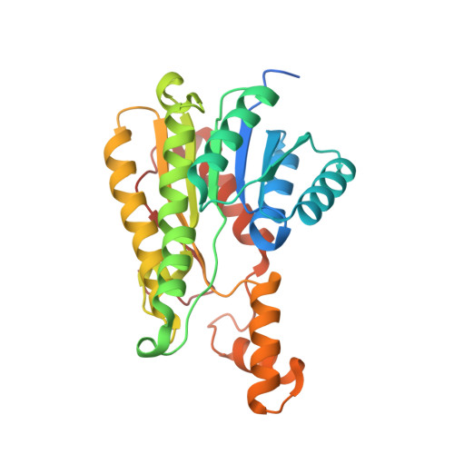
    We have determined the 2.5 angstroms crystal structure of an active, tetrameric Streptomyces coelicolor type II polyketide ketoreductase (actIII) with its bound cofactor, NADP+. This structure shows a Rossman dinucleotide binding fold characteristic of SDR enzymes. Of two subunits in the crystallographic asymmetric unit, one is closed around the active site. Formate is observed in the open subunit, indicating possible carbonyl binding sites of the polyketide intermediate. Unlike previous models we observe crystal contacts that may mimic the KR-ACP interactions that may drive active site opening. Based on these observations, we have constructed a model for ACP and polyketide binding. We propose that binding of ACP triggers a conformational change from the closed to the open, active form of the enzyme. The polyketide chain enters the active site and reduction occurs. The model also suggests a general mechanism for ACP recognition which is applicable to a range of protein families.


  • Organizational Affiliation

    Department of Biochemistry, University of Bristol, School of Medical Sciences, University Walk, Bristol, BS8 1TD, United Kingdom. a.t.hadfield@bristol.ac.uk


Macromolecules
Find similar proteins by:  (by identity cutoff)  |  3D Structure
Entity ID: 1
MoleculeChains Sequence LengthOrganismDetailsImage
KETOACYL REDUCTASE
A, B
281Streptomyces coelicolorMutation(s): 0 
EC: 1.3.1
UniProt
Find proteins for P16544 (Streptomyces coelicolor (strain ATCC BAA-471 / A3(2) / M145))
Explore P16544 
Go to UniProtKB:  P16544
Entity Groups  
Sequence Clusters30% Identity50% Identity70% Identity90% Identity95% Identity100% Identity
UniProt GroupP16544
Sequence Annotations
Expand
  • Reference Sequence
Small Molecules
Ligands 2 Unique
IDChains Name / Formula / InChI Key2D Diagram3D Interactions
NAP
Query on NAP

Download Ideal Coordinates CCD File 
C [auth A],
H [auth B]
NADP NICOTINAMIDE-ADENINE-DINUCLEOTIDE PHOSPHATE
C21 H28 N7 O17 P3
XJLXINKUBYWONI-NNYOXOHSSA-N
FMT
Query on FMT

Download Ideal Coordinates CCD File 
D [auth A]
E [auth A]
F [auth A]
G [auth A]
I [auth B]
D [auth A],
E [auth A],
F [auth A],
G [auth A],
I [auth B],
J [auth B],
K [auth B],
L [auth B],
M [auth B]
FORMIC ACID
C H2 O2
BDAGIHXWWSANSR-UHFFFAOYSA-N
Experimental Data & Validation

Experimental Data

  • Method: X-RAY DIFFRACTION
  • Resolution: 2.50 Å
  • R-Value Free: 0.218 
  • R-Value Work: 0.183 
  • R-Value Observed: 0.185 
  • Space Group: P 32 2 1
Unit Cell:
Length ( Å )Angle ( ˚ )
a = 103.43α = 90
b = 103.43β = 90
c = 122.823γ = 120
Software Package:
Software NamePurpose
REFMACrefinement
HKL-2000data reduction
SCALEPACKdata scaling
AMoREphasing

Structure Validation

View Full Validation Report


Ligand Structure Quality Assessment 


Entry History 

Deposition Data

Revision History  (Full details and data files)

  • Version 1.0: 2004-12-10
    Type: Initial release
  • Version 1.1: 2011-05-08
    Changes: Version format compliance
  • Version 1.2: 2011-07-13
    Changes: Version format compliance
  • Version 1.3: 2023-12-13
    Changes: Data collection, Database references, Other, Refinement description