1W2H

Crystal Structure Of Mycobacterium Tuberculosis Thymidylate Kinase Complexed With Azidothymidine Monophosphate (AZT-MP) (2.0 A Resolution)


Experimental Data Snapshot

  • Method: X-RAY DIFFRACTION
  • Resolution: 2.00 Å
  • R-Value Free: 0.296 
  • R-Value Work: 0.259 
  • R-Value Observed: 0.259 

wwPDB Validation   3D Report Full Report


Ligand Structure Quality Assessment 


This is version 2.1 of the entry. See complete history


Literature

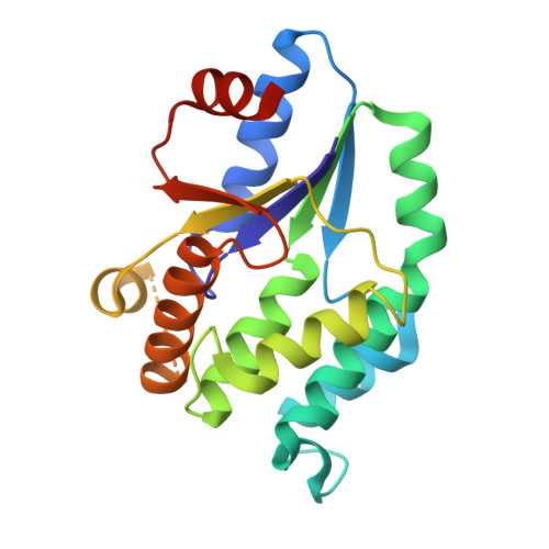
The crystal structure of Mycobacterium tuberculosis thymidylate kinase in complex with 3'-azidodeoxythymidine monophosphate suggests a mechanism for competitive inhibition.

Fioravanti, E.Adam, V.Munier-Lehmann, H.Bourgeois, D.

(2005) Biochemistry 44: 130-137

  • DOI: https://doi.org/10.1021/bi0484163
  • Primary Citation of Related Structures:  
    1W2G, 1W2H

  • PubMed Abstract: 

    Tuberculosis (TB) is the primary cause of mortality among infectious diseases. Mycobacterium tuberculosis thymidylate kinase (TMPK(Mtub)) catalyzes the ATP-dependent phosphorylation of deoxythymidine 5'-monophosphate (dTMP). Essential to DNA replication, this enzyme represents a promising target for developing new drugs against TB, because the configuration of its active site is unique within the TMPK family. Indeed, it has been proposed that, as opposed to other TMPKs, catalysis by TMPK(Mtub) necessitates the transient binding of a magnesium ion coordinating the phosphate acceptor. Moreover, 3'-azidodeoxythymidine monophosphate (AZTMP) is a competitive inhibitor of TMPK(Mtub), whereas it is a substrate for human and other TMPKs. Here, the crystal structures of TMPK(Mtub) in complex with deoxythymidine (dT) and AZTMP were determined to 2.1 and 2.0 A resolution, respectively, and suggest a mechanism for inhibition. The azido group of AZTMP perturbs the induced-fit mechanism normally adopted by the enzyme. Magnesium is prevented from binding, and the resulting electrostatic environment precludes phosphoryl transfer from occurring. Our data provide a model for drug development against tuberculosis.


  • Organizational Affiliation

    LCCP, UMR 5075, IBS-CEA/CNRS/UJF, 41 avenue Jules Horowitz, 38027 Grenoble Cedex 1, France.


Macromolecules
Find similar proteins by:  (by identity cutoff)  |  3D Structure
Entity ID: 1
MoleculeChains Sequence LengthOrganismDetailsImage
THYMIDYLATE KINASE TMK
A, B
214Mycobacterium tuberculosisMutation(s): 0 
EC: 2.7.4.9
UniProt
Find proteins for P9WKE1 (Mycobacterium tuberculosis (strain ATCC 25618 / H37Rv))
Explore P9WKE1 
Go to UniProtKB:  P9WKE1
Entity Groups  
Sequence Clusters30% Identity50% Identity70% Identity90% Identity95% Identity100% Identity
UniProt GroupP9WKE1
Sequence Annotations
Expand
  • Reference Sequence
Experimental Data & Validation

Experimental Data

  • Method: X-RAY DIFFRACTION
  • Resolution: 2.00 Å
  • R-Value Free: 0.296 
  • R-Value Work: 0.259 
  • R-Value Observed: 0.259 
  • Space Group: P 31 2 1
Unit Cell:
Length ( Å )Angle ( ˚ )
a = 64.521α = 90
b = 64.521β = 90
c = 195.366γ = 120
Software Package:
Software NamePurpose
CNSrefinement
MOSFLMdata reduction
SCALAdata scaling
CNSphasing

Structure Validation

View Full Validation Report


Ligand Structure Quality Assessment 


Entry History 

Deposition Data

Revision History  (Full details and data files)

  • Version 1.0: 2005-01-12
    Type: Initial release
  • Version 1.1: 2011-06-02
    Changes: Version format compliance
  • Version 1.2: 2011-07-13
    Changes: Version format compliance
  • Version 2.0: 2018-12-19
    Changes: Atomic model, Data collection, Database references, Derived calculations
  • Version 2.1: 2023-12-13
    Changes: Data collection, Database references, Other, Refinement description