1VZ0

Chromosome segregation protein Spo0J from Thermus thermophilus


Experimental Data Snapshot

  • Method: X-RAY DIFFRACTION
  • Resolution: 2.30 Å
  • R-Value Free: 0.278 
  • R-Value Work: 0.235 
  • R-Value Observed: 0.235 

wwPDB Validation   3D Report Full Report


This is version 1.5 of the entry. See complete history


Literature

Structural analysis of the chromosome segregation protein Spo0J from Thermus thermophilus.

Leonard, T.A.Butler, P.J.Lowe, J.

(2004) Mol Microbiol 53: 419-432

  • DOI: https://doi.org/10.1111/j.1365-2958.2004.04133.x
  • Primary Citation of Related Structures:  
    1VZ0

  • PubMed Abstract: 

    Prokaryotic chromosomes and plasmids encode partitioning systems that are required for DNA segregation at cell division. The plasmid partitioning loci encode two proteins, ParA and ParB, and a cis-acting centromere-like site denoted parS. The chromosomally encoded homologues of ParA and ParB, Soj and Spo0J, play an active role in chromosome segregation during bacterial cell division and sporulation. Spo0J is a DNA-binding protein that binds to parS sites in vivo. We have solved the X-ray crystal structure of a C-terminally truncated Spo0J (amino acids 1-222) from Thermus thermophilus to 2.3 A resolution by multiwavelength anomalous dispersion. It is a DNA-binding protein with structural similarity to the helix-turn-helix (HTH) motif of the lambda repressor DNA-binding domain. The crystal structure is an antiparallel dimer with the recognition alpha-helices of the HTH motifs of each monomer separated by a distance of 34 A corresponding to the length of the helical repeat of B-DNA. Sedimentation velocity and equilibrium ultracentrifugation studies show that full-length Spo0J exists in a monomer-dimer equilibrium in solution and that Spo0J1-222 is exclusively monomeric. Sedimentation of the C-terminal domain of Spo0J shows it to be exclusively dimeric, confirming that the C-terminus is the primary dimerization domain. We hypothesize that the C-terminus mediates dimerization of Spo0J, thereby effectively increasing the local concentration of the N-termini, which most probably dimerize, as shown by our structure, upon binding to a cognate parS site.


  • Organizational Affiliation

    MRC Laboratory of Molecular Biology, Hills Road, Cambridge CB2 2QH, UK. tleonard@mrc-lmb.cam.ac.uk


Macromolecules
Find similar proteins by:  (by identity cutoff)  |  3D Structure
Entity ID: 1
MoleculeChains Sequence LengthOrganismDetailsImage
Chromosome-partitioning protein Spo0J
A, B, C, D, E
A, B, C, D, E, F, G, H
230Thermus thermophilus HB27Mutation(s): 0 
Gene Names: spo0CparBTT_C1604
UniProt
Find proteins for Q72H91 (Thermus thermophilus (strain ATCC BAA-163 / DSM 7039 / HB27))
Explore Q72H91 
Go to UniProtKB:  Q72H91
Entity Groups  
Sequence Clusters30% Identity50% Identity70% Identity90% Identity95% Identity100% Identity
UniProt GroupQ72H91
Sequence Annotations
Expand
  • Reference Sequence
Small Molecules
Ligands 2 Unique
IDChains Name / Formula / InChI Key2D Diagram3D Interactions
CO
Query on CO

Download Ideal Coordinates CCD File 
I [auth A]
J [auth A]
K [auth A]
L [auth B]
M [auth B]
I [auth A],
J [auth A],
K [auth A],
L [auth B],
M [auth B],
N [auth C],
O [auth C],
Q [auth D],
R [auth D],
T [auth E],
U [auth E],
V [auth G],
W [auth H]
COBALT (II) ION
Co
XLJKHNWPARRRJB-UHFFFAOYSA-N
MG
Query on MG

Download Ideal Coordinates CCD File 
P [auth C],
S [auth D]
MAGNESIUM ION
Mg
JLVVSXFLKOJNIY-UHFFFAOYSA-N
Experimental Data & Validation

Experimental Data

  • Method: X-RAY DIFFRACTION
  • Resolution: 2.30 Å
  • R-Value Free: 0.278 
  • R-Value Work: 0.235 
  • R-Value Observed: 0.235 
  • Space Group: C 2 2 2
Unit Cell:
Length ( Å )Angle ( ˚ )
a = 182.24α = 90
b = 295.15β = 90
c = 73.49γ = 90
Software Package:
Software NamePurpose
CNSrefinement
MOSFLMdata reduction
SCALAdata scaling
SnBphasing

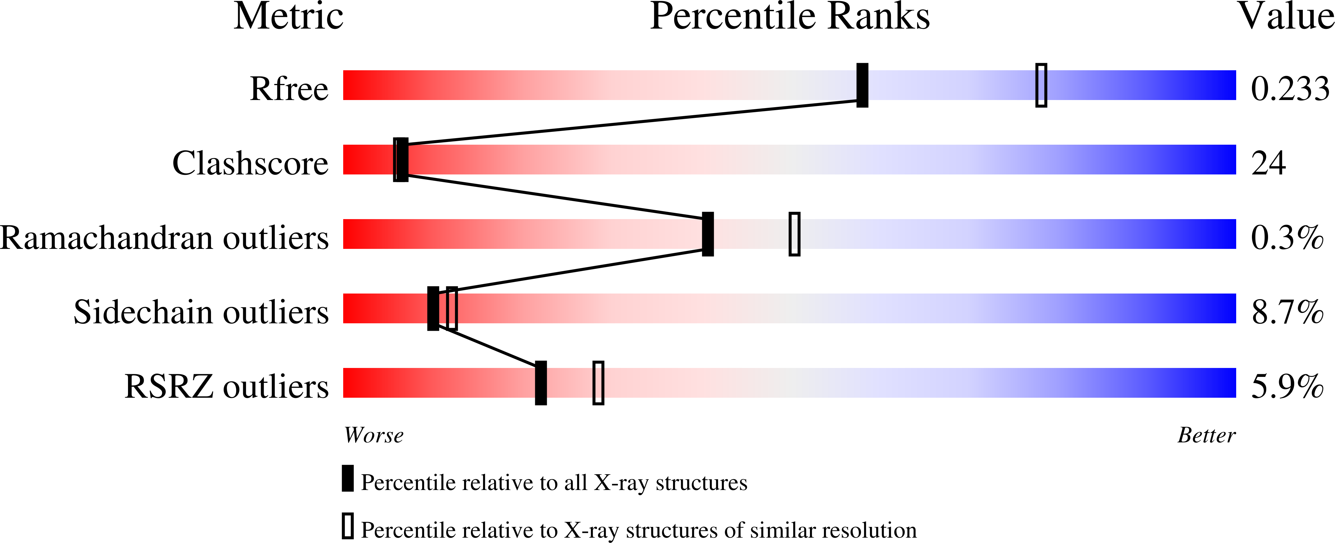
Structure Validation

View Full Validation Report


Entry History 

Deposition Data

Revision History  (Full details and data files)

  • Version 1.0: 2004-07-19
    Type: Initial release
  • Version 1.1: 2011-05-08
    Changes: Version format compliance
  • Version 1.2: 2011-07-13
    Changes: Version format compliance
  • Version 1.3: 2017-07-05
    Changes: Refinement description
  • Version 1.4: 2018-01-24
    Changes: Source and taxonomy
  • Version 1.5: 2018-06-20
    Changes: Data collection, Database references, Source and taxonomy, Structure summary