1VYZ

Structure of CDK2 complexed with PNU-181227


Experimental Data Snapshot

  • Method: X-RAY DIFFRACTION
  • Resolution: 2.21 Å
  • R-Value Free: 0.280 
  • R-Value Work: 0.238 

wwPDB Validation   3D Report Full Report


Ligand Structure Quality Assessment 


This is version 1.3 of the entry. See complete history


Literature

3-Aminopyrazole Inhibitors of Cdk2/Cyclin a as Antitumor Agents. 1. Lead Finding

Pevarello, P.Brasca, M.G.Amici, R.Orsini, P.Traquandi, G.Corti, L.Piutti, C.Sansonna, P.Villa, M.Pierce, B.S.Pulici, M.Giordano, P.Martina, K.Fritzen, E.Nugent, R.A.Casale, E.Cameron, A.Ciomei, M.Roletto, F.Isacchi, A.Fogliatto, G.Pesenti, E.Pastori, W.Marsiglio, A.Leach, K.L.Clare, P.M.Fiorentini, F.Varasi, M.Vulpetti, A.Warpehoski, M.A.

(2004) J Med Chem 47: 3367

  • DOI: https://doi.org/10.1021/jm031145u
  • Primary Citation of Related Structures:  
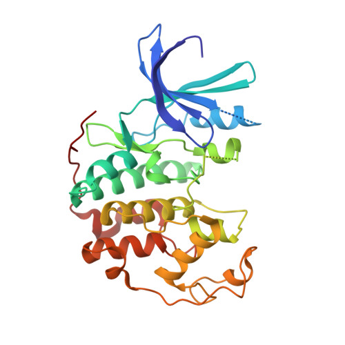
    1VYW, 1VYZ

  • PubMed Abstract: 

    Abnormal proliferation mediated by disruption of the normal cell cycle mechanisms is a hallmark of virtually all cancer cells. Compounds targeting complexes between cyclin-dependent kinases (CDK) and cyclins, such as CDK2/cyclin A and CDK2/cyclin E, and inhibiting their kinase activity are regarded as promising antitumor agents to complement the existing therapies. From a high-throughput screening effort, we identified a new class of CDK2/cyclin A/E inhibitors. The hit-to-lead expansion of this class is described. X-ray crystallographic data of early compounds in this series, as well as in vitro testing funneled for rapidly achieving in vivo efficacy, led to a nanomolar inhibitor of CDK2/cyclin A (N-(5-cyclopropyl-1H-pyrazol-3-yl)-2-(2-naphthyl)acetamide (41), PNU-292137, IC50 = 37 nM) with in vivo antitumor activity (TGI > 50%) in a mouse xenograft model at a dose devoid of toxic effects.


  • Organizational Affiliation

    Chemistry Department, Pharmacia Italia, Viale Pasteur 10, 20014 Nerviano (MI), Italy. paolo.pevarello@pharmacia.com


Macromolecules
Find similar proteins by:  (by identity cutoff)  |  3D Structure
Entity ID: 1
MoleculeChains Sequence LengthOrganismDetailsImage
CELL DIVISION PROTEIN KINASE 2298Homo sapiensMutation(s): 0 
EC: 2.7.1.37
UniProt & NIH Common Fund Data Resources
Find proteins for P24941 (Homo sapiens)
Explore P24941 
Go to UniProtKB:  P24941
PHAROS:  P24941
GTEx:  ENSG00000123374 
Entity Groups  
Sequence Clusters30% Identity50% Identity70% Identity90% Identity95% Identity100% Identity
UniProt GroupP24941
Sequence Annotations
Expand
  • Reference Sequence
Small Molecules
Ligands 1 Unique
IDChains Name / Formula / InChI Key2D Diagram3D Interactions
N5B
Query on N5B

Download Ideal Coordinates CCD File 
B [auth A]N-(5-CYCLOPROPYL-1H-PYRAZOL-3-YL)BENZAMIDE
C13 H13 N3 O
LUCORKWTQSQFFU-UHFFFAOYSA-N
Binding Affinity Annotations 
IDSourceBinding Affinity
N5B BindingDB:  1VYZ IC50: 290 (nM) from 1 assay(s)
PDBBind:  1VYZ IC50: 290 (nM) from 1 assay(s)
Binding MOAD:  1VYZ IC50: 290 (nM) from 1 assay(s)
Experimental Data & Validation

Experimental Data

  • Method: X-RAY DIFFRACTION
  • Resolution: 2.21 Å
  • R-Value Free: 0.280 
  • R-Value Work: 0.238 
  • Space Group: P 21 21 21
Unit Cell:
Length ( Å )Angle ( ˚ )
a = 53.25α = 90
b = 71.91β = 90
c = 72.22γ = 90
Software Package:
Software NamePurpose
CNXrefinement
SCALEPACKdata scaling

Structure Validation

View Full Validation Report


Ligand Structure Quality Assessment 


Entry History 

Revision History  (Full details and data files)

  • Version 1.0: 2004-06-17
    Type: Initial release
  • Version 1.1: 2011-05-08
    Changes: Version format compliance
  • Version 1.2: 2011-07-13
    Changes: Version format compliance
  • Version 1.3: 2019-04-03
    Changes: Data collection, Other, Source and taxonomy