1VYB

Endonuclease domain of human LINE1 ORF2p


Experimental Data Snapshot

  • Method: X-RAY DIFFRACTION
  • Resolution: 1.80 Å
  • R-Value Free: 0.223 
  • R-Value Work: 0.186 
  • R-Value Observed: 0.188 

wwPDB Validation   3D Report Full Report


This is version 1.4 of the entry. See complete history


Literature

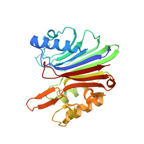
Crystal structure of the targeting endonuclease of the human LINE-1 retrotransposon.

Weichenrieder, O.Repanas, K.Perrakis, A.

(2004) Structure 12: 975-986

  • DOI: https://doi.org/10.1016/j.str.2004.04.011
  • Primary Citation of Related Structures:  
    1VYB

  • PubMed Abstract: 

    The human L1 endonuclease (L1-EN) is encoded by the non-LTR retrotransposon LINE-1 (L1). L1 is responsible for more than 1.5 million retrotransposition events in the history of the human genome, contributing more than a quarter to human genomic DNA (L1 and Alu elements). L1-EN is related to the well-understood human DNA repair endonuclease APE1, and its nicking specificity is a major determinant for retrotransposon integration site selection. The crystal structure of human L1 endonuclease is the first of a retrotransposon-encoded protein and a prototype for retrotransposon-encoded endonucleases involved in target-primed reverse transcription. Structure-based endonuclease alignments reveal a conserved threonine in addition to previously identified invariant residues and suggest that DNA recognition proceeds via the accommodation of an extrahelical nucleotide within a pocket of the enzyme. The present analysis will help to refine phylogenetic and functional relationships among metal-dependent phosphohydrolases and provides a basis for manipulating non-LTR retrotransposon integration site selection.


  • Organizational Affiliation

    The Netherlands Cancer Institute, Department of Molecular Carcinogenesis-H2, Plesmanlaan 121, 1066 CX Amsterdam, The Netherlands. o.weichenrieder@nki.nl


Macromolecules
Find similar proteins by:  (by identity cutoff)  |  3D Structure
Entity ID: 1
MoleculeChains Sequence LengthOrganismDetailsImage
ORF2 CONTAINS A REVERSE TRANSCRIPTASE DOMAIN
A, B
238Homo sapiensMutation(s): 0 
UniProt
Find proteins for O00370 (Homo sapiens)
Explore O00370 
Go to UniProtKB:  O00370
Entity Groups  
Sequence Clusters30% Identity50% Identity70% Identity90% Identity95% Identity100% Identity
UniProt GroupO00370
Sequence Annotations
Expand
  • Reference Sequence
Small Molecules
Ligands 3 Unique
IDChains Name / Formula / InChI Key2D Diagram3D Interactions
SO4
Query on SO4

Download Ideal Coordinates CCD File 
C [auth A]
D [auth A]
E [auth A]
H [auth B]
I [auth B]
C [auth A],
D [auth A],
E [auth A],
H [auth B],
I [auth B],
J [auth B],
K [auth B]
SULFATE ION
O4 S
QAOWNCQODCNURD-UHFFFAOYSA-L
GOL
Query on GOL

Download Ideal Coordinates CCD File 
G [auth A]GLYCEROL
C3 H8 O3
PEDCQBHIVMGVHV-UHFFFAOYSA-N
SO3
Query on SO3

Download Ideal Coordinates CCD File 
F [auth A],
L [auth B]
SULFITE ION
O3 S
LSNNMFCWUKXFEE-UHFFFAOYSA-L
Experimental Data & Validation

Experimental Data

  • Method: X-RAY DIFFRACTION
  • Resolution: 1.80 Å
  • R-Value Free: 0.223 
  • R-Value Work: 0.186 
  • R-Value Observed: 0.188 
  • Space Group: P 21 21 2
Unit Cell:
Length ( Å )Angle ( ˚ )
a = 91.044α = 90
b = 126.477β = 90
c = 43.004γ = 90
Software Package:
Software NamePurpose
REFMACrefinement
DENZOdata reduction
SCALEPACKdata scaling
AMoREphasing

Structure Validation

View Full Validation Report


Entry History 

Deposition Data

Revision History  (Full details and data files)

  • Version 1.0: 2004-06-04
    Type: Initial release
  • Version 1.1: 2011-05-08
    Changes: Version format compliance
  • Version 1.2: 2011-07-13
    Changes: Version format compliance
  • Version 1.3: 2018-02-28
    Changes: Database references, Source and taxonomy
  • Version 1.4: 2023-12-13
    Changes: Data collection, Database references, Other, Refinement description