1VSO

Crystal Structure of the Ligand-Binding Core of iGluR5 in Complex With the Antagonist (S)-ATPO at 1.85 A resolution


Experimental Data Snapshot

  • Method: X-RAY DIFFRACTION
  • Resolution: 1.85 Å
  • R-Value Free: 0.231 
  • R-Value Work: 0.202 
  • R-Value Observed: 0.202 

wwPDB Validation   3D Report Full Report


Ligand Structure Quality Assessment 


This is version 1.5 of the entry. See complete history


Literature

Partial agonism and antagonism of the ionotropic glutamate receptor iGLuR5: structures of the ligand-binding core in complex with domoic acid and 2-amino-3-[5-tert-butyl-3-(phosphonomethoxy)-4-isoxazolyl]propionic acid.

Hald, H.Naur, P.Pickering, D.S.Sprogoe, D.Madsen, U.Timmermann, D.B.Ahring, P.K.Liljefors, T.Schousboe, A.Egebjerg, J.Gajhede, M.Kastrup, J.S.

(2007) J Biol Chem 282: 25726-25736

  • DOI: https://doi.org/10.1074/jbc.M700137200
  • Primary Citation of Related Structures:  
    1VSO, 2PBW

  • PubMed Abstract: 

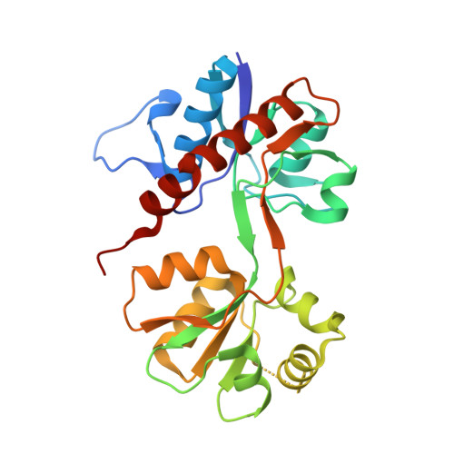
    More than 50 structures have been reported on the ligand-binding core of the ionotropic glutamate receptor iGluR2 that belongs to the 2-amino-3-(3-hydroxy-5-methyl-4-isoxazolyl)propionic acid-type of receptors. In contrast, the ligand-binding core of the kainic acid-type receptor iGluR5 has only been crystallized with three different ligands. Hence, additional structures of iGluR5 are needed to broaden the understanding of the ligand-binding properties of iGluR5, and the conformational changes leading to channel opening and closing. Here, we present two structures of the ligand-binding core of iGluR5; one as a complex with the partial agonist (2S,3S,4S)-3-carboxymethyl-4-[(1Z,3E,5R)-5-carboxy-1-methyl-hexa-1,3-dienyl]-pyrrolidine-2-carboxylic acid (domoic acid) and one as a complex with the antagonist (S)-2-amino-3-[5-tert-butyl-3-(phosphonomethoxy)-4-isoxazolyl]propionic acid ((S)-ATPO). In agreement with the partial agonist activity of domoic acid, the ligand-binding core of the iGluR5 complex is stabilized by domoic acid in a conformation that is 11 degrees more open than the conformation observed in the full agonist (S)-glutamic acid complex. This is primarily caused by the 5-carboxy-1-methyl-hexa-1,3-dienyl moiety of domoic acid and residues Val685-Thr690 of iGluR5. An even larger domain opening of 28 degrees is introduced upon binding of the antagonist (S)-ATPO. It appears that the span of domain opening is much larger in the ligand-binding core of iGluR5 (30 degrees) compared with what has been observed in iGluR2 (19 degrees ). Similarly, much larger variation in the distances between transmembrane linker residues in the two protomers comprising the dimer is observed in iGluR5 as compared with iGluR2.


  • Organizational Affiliation

    Department of Medicinal Chemistry, Faculty of Pharmaceutical Sciences, University of Copenhagen, Universitetsparken 2, DK-2100, Copenhagen, Denmark.


Macromolecules
Find similar proteins by:  (by identity cutoff)  |  3D Structure
Entity ID: 1
MoleculeChains Sequence LengthOrganismDetailsImage
Glutamate receptor, ionotropic kainate 1257Rattus norvegicusMutation(s): 0 
Gene Names: Grik1Glur5
UniProt
Find proteins for P22756 (Rattus norvegicus)
Explore P22756 
Go to UniProtKB:  P22756
Entity Groups  
Sequence Clusters30% Identity50% Identity70% Identity90% Identity95% Identity100% Identity
UniProt GroupP22756
Sequence Annotations
Expand
  • Reference Sequence
Small Molecules
Ligands 2 Unique
IDChains Name / Formula / InChI Key2D Diagram3D Interactions
AT1
Query on AT1

Download Ideal Coordinates CCD File 
B [auth A](S)-2-AMINO-3-(5-TERT-BUTYL-3-(PHOSPHONOMETHOXY)-4-ISOXAZOLYL)PROPIONIC ACID
C11 H19 N2 O7 P
AGSOOCUNMTYPSE-ZETCQYMHSA-N
GOL
Query on GOL

Download Ideal Coordinates CCD File 
C [auth A]GLYCEROL
C3 H8 O3
PEDCQBHIVMGVHV-UHFFFAOYSA-N
Binding Affinity Annotations 
IDSourceBinding Affinity
AT1 Binding MOAD:  1VSO Ki: 1.90e+4 (nM) from 1 assay(s)
PDBBind:  1VSO Ki: 1.90e+4 (nM) from 1 assay(s)
BindingDB:  1VSO Ki: 2900 (nM) from 1 assay(s)
Experimental Data & Validation

Experimental Data

  • Method: X-RAY DIFFRACTION
  • Resolution: 1.85 Å
  • R-Value Free: 0.231 
  • R-Value Work: 0.202 
  • R-Value Observed: 0.202 
  • Space Group: P 64
Unit Cell:
Length ( Å )Angle ( ˚ )
a = 108.9α = 90
b = 108.9β = 90
c = 51.092γ = 120
Software Package:
Software NamePurpose
DENZOdata reduction
SCALEPACKdata scaling
PHASERphasing
CNSrefinement
PDB_EXTRACTdata extraction
HKL-2000data reduction
HKL-2000data scaling

Structure Validation

View Full Validation Report


Ligand Structure Quality Assessment 


Entry History 

Deposition Data

Revision History  (Full details and data files)

  • Version 1.0: 2007-07-03
    Type: Initial release
  • Version 1.1: 2007-10-03
    Changes: Version format compliance
  • Version 1.2: 2011-07-13
    Changes: Non-polymer description, Version format compliance
  • Version 1.3: 2017-08-02
    Changes: Refinement description, Source and taxonomy
  • Version 1.4: 2017-10-04
    Changes: Refinement description
  • Version 1.5: 2023-08-23
    Changes: Data collection, Database references, Derived calculations, Refinement description