1VSN

Crystal structure of a potent small molecule inhibitor bound to cathepsin K


Experimental Data Snapshot

  • Method: X-RAY DIFFRACTION
  • Resolution: 2.00 Å
  • R-Value Free: 0.209 
  • R-Value Work: 0.171 
  • R-Value Observed: 0.212 

wwPDB Validation   3D Report Full Report


Ligand Structure Quality Assessment 


This is version 1.5 of the entry. See complete history


Literature

Identification of a potent and selective non-basic cathepsin K inhibitor.

Li, C.S.Deschenes, D.Desmarais, S.Falgueyret, J.P.Gauthier, J.Y.Kimmel, D.B.McGrath, M.E.McKay, D.J.Percival, M.D.Riendeau, D.Rodan, S.B.Truong, V.L.Wesolowski, G.Zamboni, R.Black, W.C.

(2006) Bioorg Med Chem Lett 16: 1985-1989

  • DOI: https://doi.org/10.1016/j.bmcl.2005.12.071
  • Primary Citation of Related Structures:  
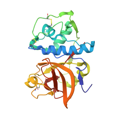
    1VSN

  • PubMed Abstract: 

    Based on our previous study with trifluoroethylamine as a P2-P3 amide isostere of cathepsin K inhibitor, further optimization led to identification of compound 22 (L-873724) as a potent and selective non-basic cathepsin K inhibitor. This compound showed excellent pharmacokinetics and efficacy in an ovariectomized (OVX) rhesus monkey model. The volumes of distribution close to unity were consistent with this compound not being lysosomotropic, which is a characteristic of basic cathepsin K inhibitors.


  • Organizational Affiliation

    Merck Frosst Centre for Therapeutic Research, PO Box 1005, Pointe-Claire-Dorval, Que., Canada H9R 4P8. chunsing_li@merck.com


Macromolecules
Find similar proteins by:  (by identity cutoff)  |  3D Structure
Entity ID: 1
MoleculeChains Sequence LengthOrganismDetailsImage
Cathepsin K215Homo sapiensMutation(s): 0 
Gene Names: CTSKCTSOCTSO2
EC: 3.4.22.38
UniProt & NIH Common Fund Data Resources
Find proteins for P43235 (Homo sapiens)
Explore P43235 
Go to UniProtKB:  P43235
PHAROS:  P43235
GTEx:  ENSG00000143387 
Entity Groups  
Sequence Clusters30% Identity50% Identity70% Identity90% Identity95% Identity100% Identity
UniProt GroupP43235
Sequence Annotations
Expand
  • Reference Sequence
Small Molecules
Ligands 1 Unique
IDChains Name / Formula / InChI Key2D Diagram3D Interactions
NFT
Query on NFT

Download Ideal Coordinates CCD File 
B [auth A]N-(2-AMINOETHYL)-N~2~-{(1S)-1-[4'-(AMINOSULFONYL)BIPHENYL-4-YL]-2,2,2-TRIFLUOROETHYL}-L-LEUCINAMIDE
C22 H29 F3 N4 O3 S
QPXXBNKMAHUXBB-PMACEKPBSA-N
Binding Affinity Annotations 
IDSourceBinding Affinity
NFT PDBBind:  1VSN IC50: 0.2 (nM) from 1 assay(s)
Experimental Data & Validation

Experimental Data

  • Method: X-RAY DIFFRACTION
  • Resolution: 2.00 Å
  • R-Value Free: 0.209 
  • R-Value Work: 0.171 
  • R-Value Observed: 0.212 
  • Space Group: P 21 21 21
Unit Cell:
Length ( Å )Angle ( ˚ )
a = 38.42α = 90
b = 51β = 90
c = 104.2γ = 90
Software Package:
Software NamePurpose
CNSrefinement
REFMACrefinement
PDB_EXTRACTdata extraction
bioteXdata reduction
XTALVIEWrefinement
d*TREKdata scaling

Structure Validation

View Full Validation Report


Ligand Structure Quality Assessment 


Entry History 

Deposition Data

  • Released Date: 2007-04-24 
  • Deposition Author(s): McGrath, M.
  • This entry supersedes: 2FDZ

Revision History  (Full details and data files)

  • Version 1.0: 2007-04-24
    Type: Initial release
  • Version 1.1: 2008-04-27
    Changes: Version format compliance
  • Version 1.2: 2011-07-13
    Changes: Version format compliance
  • Version 1.3: 2017-10-04
    Changes: Refinement description
  • Version 1.4: 2019-07-24
    Changes: Data collection, Derived calculations, Refinement description
  • Version 1.5: 2023-12-27
    Changes: Data collection, Database references, Derived calculations