1VR1

Specifity for Plasminogen Activator Inhibitor-1


Experimental Data Snapshot

  • Method: X-RAY DIFFRACTION
  • Resolution: 1.90 Å
  • R-Value Free: 0.256 
  • R-Value Work: 0.204 

wwPDB Validation   3D Report Full Report


This is version 1.5 of the entry. See complete history


Literature

The variable region-1 from tissue-type plasminogen activator confers specificity for plasminogen activator inhibitor-1 to thrombin by facilitating catalysis: release of a kinetic block by a heterologous protein surface loop

Dekker, R.J.Eichinger, A.Stoop, A.A.Bode, W.Pannekoek, H.Horrevoets, A.J.G.

(1999) J Mol Biol 293: 613-627

  • DOI: https://doi.org/10.1006/jmbi.1999.3178
  • Primary Citation of Related Structures:  
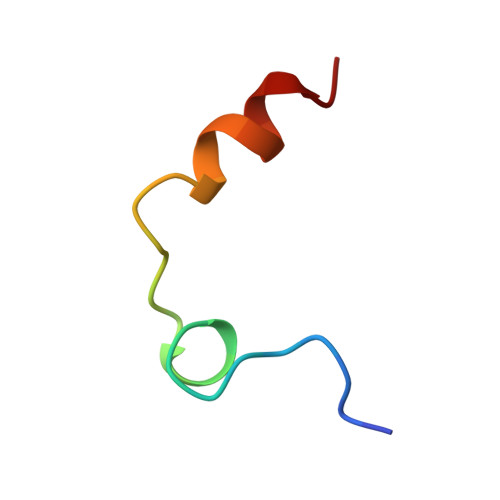
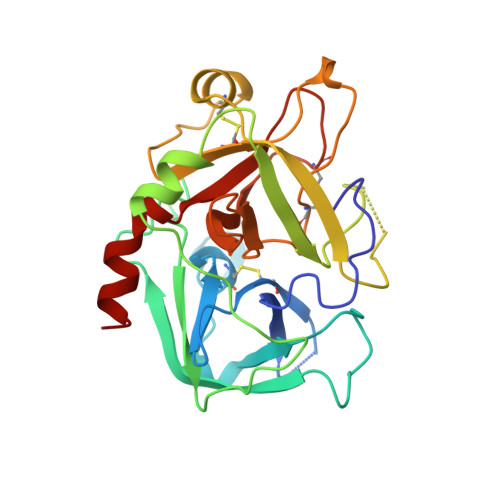
    1VR1

  • PubMed Abstract: 

    Substitution of the native variable region-1 (VR1/37-loop) of thrombin by the corresponding VR1 of tissue-type plasminogen activator (thrombin-VR1(tPA)) increases the rate of inhibition by plasminogen activator inhibitor type 1 (PAI-1) by three orders of magnitude, and is thus sufficient to confer PAI-1 specificity to a heterologous serine protease. A structural and kinetical approach to establish the function of the VR1 loop of t-PA in the context of the thrombin-VR1(tPA) variant is described. The crystal structure of thrombin-VR1(tPA) was resolved and showed a conserved overall alpha-thrombin structure, but a partially disordered VR1 loop as also reported for t-PA. The contribution of a prominent charge substitution close to the active site was studied using charge neutralization variants thrombin-E39Q(c39) and thrombin-VR1(tPA)-R304Q(c39), resulting in only fourfold changes in the PAI-1 inhibition rate. Surface plasmon resonance revealed that the affinity of initial reversible complex formation between PAI-1 and catalytically inactive Ser195-->Ala variants of thrombin and thrombin-VR1(tPA) is only increased fivefold, i.e. KD is 652 and 128 nM for thrombin-S195A and thrombin-S195A-VR1(tPA), respectively. We established that the partition ratio of the suicide substrate reaction between the proteases and PAI-1 was largely unaffected in any variant studied. Hirugen allosterically decreases the rate of thrombin inhibition by PAI-1 2.5-fold and of thrombin-VR1(tPA) 20-fold, by interfering with a unimolecular step in the reaction, not by decreasing initial complex formation or by altering the stoichiometry. Finally, kinetic modeling demonstrated that acylation is the rate-limiting step in thrombin inhibition by PAI-1 (k approximately 10(-3) s(-1)) and this kinetic block is alleviated by the introduction of the tPA-VR1 into thrombin (k>1 s(-1)). We propose that the length, flexibility and different charge architecture of the VR1 loop of t-PA invoke an induced fit of the reactive center loop of PAI-1, thereby enhancing the rate of acylation in the Michaelis complex between thrombin-VR1(t-PA) and PAI-1 by more than two orders of magnitude.


  • Organizational Affiliation

    Department of Biochemistry Academic Medical Center, University of Amsterdam, Amsterdam, 1105 AZ, The Netherlands.


Macromolecules
Find similar proteins by:  (by identity cutoff)  |  3D Structure
Entity ID: 1
MoleculeChains Sequence LengthOrganismDetailsImage
thrombinA [auth L]27Homo sapiensMutation(s): 0 
Gene Names: F2
EC: 3.4.21.5
UniProt & NIH Common Fund Data Resources
Find proteins for P00734 (Homo sapiens)
Explore P00734 
Go to UniProtKB:  P00734
PHAROS:  P00734
GTEx:  ENSG00000180210 
Entity Groups  
Sequence Clusters30% Identity50% Identity70% Identity90% Identity95% Identity100% Identity
UniProt GroupP00734
Sequence Annotations
Expand
  • Reference Sequence
Find similar proteins by:  (by identity cutoff)  |  3D Structure
Entity ID: 2
MoleculeChains Sequence LengthOrganismDetailsImage
thrombinB [auth H]261Homo sapiensMutation(s): 0 
Gene Names: F2
EC: 3.4.21.5
UniProt & NIH Common Fund Data Resources
Find proteins for P00734 (Homo sapiens)
Explore P00734 
Go to UniProtKB:  P00734
PHAROS:  P00734
GTEx:  ENSG00000180210 
Entity Groups  
Sequence Clusters30% Identity50% Identity70% Identity90% Identity95% Identity100% Identity
UniProt GroupP00734
Sequence Annotations
Expand
  • Reference Sequence

Find similar proteins by:  Sequence   |   3D Structure  

Entity ID: 3
MoleculeChains Sequence LengthOrganismDetailsImage
HirudinC [auth I]10Hirudo medicinalisMutation(s): 0 
UniProt
Find proteins for P01050 (Hirudo medicinalis)
Explore P01050 
Go to UniProtKB:  P01050
Entity Groups  
Sequence Clusters30% Identity50% Identity70% Identity90% Identity95% Identity100% Identity
UniProt GroupP01050
Sequence Annotations
Expand
  • Reference Sequence
Small Molecules
Modified Residues  1 Unique
IDChains TypeFormula2D DiagramParent
TYS
Query on TYS
C [auth I]L-PEPTIDE LINKINGC9 H11 N O6 STYR
Experimental Data & Validation

Experimental Data

  • Method: X-RAY DIFFRACTION
  • Resolution: 1.90 Å
  • R-Value Free: 0.256 
  • R-Value Work: 0.204 
  • Space Group: C 1 2 1
Unit Cell:
Length ( Å )Angle ( ˚ )
a = 70.88α = 90
b = 71.96β = 100.65
c = 73.04γ = 90
Software Package:
Software NamePurpose
REFMACrefinement

Structure Validation

View Full Validation Report


Entry History 

Deposition Data

Revision History  (Full details and data files)

  • Version 1.0: 1999-12-29
    Type: Initial release
  • Version 1.1: 2008-04-26
    Changes: Version format compliance
  • Version 1.2: 2011-07-13
    Changes: Atomic model, Database references, Derived calculations, Non-polymer description, Structure summary, Version format compliance
  • Version 1.3: 2012-12-12
    Changes: Other
  • Version 1.4: 2013-03-13
    Changes: Other
  • Version 1.5: 2022-12-21
    Changes: Database references, Derived calculations