1VLN

A TRICLINIC CRYSTAL FORM OF THE LECTIN CONCANAVALIN A


Experimental Data Snapshot

  • Method: X-RAY DIFFRACTION
  • Resolution: 2.40 Å
  • R-Value Free: 0.265 
  • R-Value Work: 0.205 
  • R-Value Observed: 0.205 

wwPDB Validation   3D Report Full Report


This is version 1.3 of the entry. See complete history


Literature

A Triclinic Crystal Form of the Lectin Concanavalin A

Kanellopoulos, P.N.Tucker, P.A.Pavlou, K.Agianian, B.Hamodrakas, S.J.

(1996) J Struct Biol 117: 16-23

  • DOI: https://doi.org/10.1006/jsbi.1996.0065
  • Primary Citation of Related Structures:  
    1VLN

  • PubMed Abstract: 

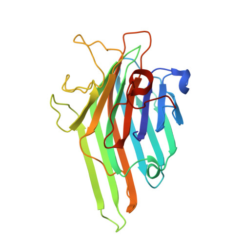
    The molecular structure of a triclinic crystal form of concanavalin A has been refined at 2.4 A resolution. The crystals have unit cell dimensions a = 78.8 A, b = 79.3 A, c = 133.3 A, alpha = 97.1degrees, beta = 90.2degrees, and gamma = 97.5degrees and contain two tetramers per asymmetric unit each with approximate 222 symmetry. The final crystallographic R-factor is 0.205 and the free-R-factor is 0.265 in the resolution range 6.0 to 2.4 A. The conformation of the tetramer is more similar to that found in concanavalin A saccharide complexes than in the previously reported I222 crystal form of uncomplexed concanavalin A. A comparison of the molecular packing between the two crystal forms shows a more open arrangement with large solvent channels through the crystal.


  • Organizational Affiliation

    European Molecular Biology Laboratory, Heidelberg, D-69012, Germany


Macromolecules
Find similar proteins by:  (by identity cutoff)  |  3D Structure
Entity ID: 1
MoleculeChains Sequence LengthOrganismDetailsImage
CONCANAVALIN A
A, B, C, D, E
A, B, C, D, E, F, G, H
237Canavalia ensiformisMutation(s): 0 
UniProt
Find proteins for P08902 (Dioclea grandiflora)
Explore P08902 
Go to UniProtKB:  P08902
Entity Groups  
Sequence Clusters30% Identity50% Identity70% Identity90% Identity95% Identity100% Identity
UniProt GroupP08902
Sequence Annotations
Expand
  • Reference Sequence
Small Molecules
Ligands 2 Unique
IDChains Name / Formula / InChI Key2D Diagram3D Interactions
MN
Query on MN

Download Ideal Coordinates CCD File 
I [auth A]
K [auth B]
M [auth C]
O [auth D]
Q [auth E]
I [auth A],
K [auth B],
M [auth C],
O [auth D],
Q [auth E],
S [auth F],
U [auth G],
W [auth H]
MANGANESE (II) ION
Mn
WAEMQWOKJMHJLA-UHFFFAOYSA-N
CA
Query on CA

Download Ideal Coordinates CCD File 
J [auth A]
L [auth B]
N [auth C]
P [auth D]
R [auth E]
J [auth A],
L [auth B],
N [auth C],
P [auth D],
R [auth E],
T [auth F],
V [auth G],
X [auth H]
CALCIUM ION
Ca
BHPQYMZQTOCNFJ-UHFFFAOYSA-N
Experimental Data & Validation

Experimental Data

  • Method: X-RAY DIFFRACTION
  • Resolution: 2.40 Å
  • R-Value Free: 0.265 
  • R-Value Work: 0.205 
  • R-Value Observed: 0.205 
  • Space Group: P 1
Unit Cell:
Length ( Å )Angle ( ˚ )
a = 78.8α = 97.1
b = 79.3β = 90.2
c = 133.3γ = 97.5
Software Package:
Software NamePurpose
X-PLORmodel building
X-PLORrefinement
XDSdata reduction
XSCALEdata scaling
X-PLORphasing

Structure Validation

View Full Validation Report


Entry History 

Deposition Data

Revision History  (Full details and data files)

  • Version 1.0: 1997-04-01
    Type: Initial release
  • Version 1.1: 2008-03-24
    Changes: Version format compliance
  • Version 1.2: 2011-07-13
    Changes: Version format compliance
  • Version 1.3: 2023-08-09
    Changes: Database references, Derived calculations, Other, Refinement description