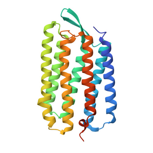
1VJM

Deformation of helix C in the low-temperature L-intermediate of bacteriorhodopsin


Experimental Data Snapshot

  • Method: X-RAY DIFFRACTION
  • Resolution: 2.30 Å
  • R-Value Free: 0.298 
  • R-Value Work: 0.252 
  • R-Value Observed: 0.257 

wwPDB Validation   3D Report Full Report


Ligand Structure Quality Assessment 


This is version 1.3 of the entry. See complete history


Literature

Deformation of helix C in the low temperature L-intermediate of bacteriorhodopsin.

Edman, K.Royant, A.Larsson, G.Jacobson, F.Taylor, T.van der Spoel, D.Landau, E.M.Pebay-Peyroula, E.Neutze, R.

(2004) J Biol Chem 279: 2147-2158

  • DOI: https://doi.org/10.1074/jbc.M300709200
  • Primary Citation of Related Structures:  
    1VJM

  • PubMed Abstract: 

    X-ray and electron diffraction studies of specific reaction intermediates, or reaction intermediate analogues, have produced a consistent picture of the structural mechanism of light-driven proton pumping by bacteriorhodopsin. Of central importance within this picture is the structure of the L-intermediate, which follows the retinal all-trans to 13-cis photoisomerization step of the K-intermediate and sets the stage for the primary proton transfer event from the positively charged Schiff base to the negatively charged Asp-85. Here we report the structural changes in bacteriorhodopsin following red light illumination at 150 K. Single crystal microspectrophotometry showed that only the L-intermediate is populated in three-dimensional crystals under these conditions. The experimental difference Fourier electron density map and refined crystallographic structure were consistent with those previously presented (Royant, A., Edman, K., Ursby, T., Pebay-Peyroula, E., Landau, E. M., and Neutze, R. (2000) Nature 406, 645-648; Royant, A., Edman, K., Ursby, T., Pebay-Peyroula, E., Landau, E. M., and Neutze, R. (2001) Photochem. Photobiol. 74, 794-804). Based on the refined crystallographic structures, molecular dynamic simulations were used to examine the influence of the conformational change of the protein that is associated with the K-to-L transition on retinal dynamics. Implications regarding the structural mechanism for proton pumping by bacteriorhodopsin are discussed.


  • Organizational Affiliation

    Department of Chemistry and Bioscience, Chalmers University of Technology, Box 462, S-40530 Gothenburg, Sweden.


Macromolecules
Find similar proteins by:  (by identity cutoff)  |  3D Structure
Entity ID: 1
MoleculeChains Sequence LengthOrganismDetailsImage
Bacteriorhodopsin249Halobacterium sp.Mutation(s): 0 
Membrane Entity: Yes 
UniProt
Find proteins for P02945 (Halobacterium salinarum (strain ATCC 700922 / JCM 11081 / NRC-1))
Explore P02945 
Go to UniProtKB:  P02945
Entity Groups  
Sequence Clusters30% Identity50% Identity70% Identity90% Identity95% Identity100% Identity
UniProt GroupP02945
Sequence Annotations
Expand
  • Reference Sequence
Small Molecules
Ligands 1 Unique
IDChains Name / Formula / InChI Key2D Diagram3D Interactions
RET
Query on RET

Download Ideal Coordinates CCD File 
B [auth A]RETINAL
C20 H28 O
NCYCYZXNIZJOKI-OVSJKPMPSA-N
Experimental Data & Validation

Experimental Data

  • Method: X-RAY DIFFRACTION
  • Resolution: 2.30 Å
  • R-Value Free: 0.298 
  • R-Value Work: 0.252 
  • R-Value Observed: 0.257 
  • Space Group: P 63
Unit Cell:
Length ( Å )Angle ( ˚ )
a = 60.87α = 90
b = 60.87β = 90
c = 109.5γ = 120
Software Package:
Software NamePurpose
DENZOdata reduction
SCALAdata scaling
AMoREphasing
CNSrefinement
CCP4data scaling

Structure Validation

View Full Validation Report


Ligand Structure Quality Assessment 


Entry History 

Deposition Data

Revision History  (Full details and data files)

  • Version 1.0: 2004-04-06
    Type: Initial release
  • Version 1.1: 2008-04-26
    Changes: Version format compliance
  • Version 1.2: 2011-07-13
    Changes: Derived calculations, Version format compliance
  • Version 1.3: 2023-10-25
    Changes: Data collection, Database references, Derived calculations, Refinement description