Structural studies on bioactive compounds. 30. Crystal structure and molecular modeling studies on the Pneumocystis carinii dihydrofolate reductase cofactor complex with TAB, a highly selective antifolate.
Cody, V., Chan, D., Galitsky, N., Rak, D., Luft, J.R., Pangborn, W., Queener, S.F., Laughton, C.A., Stevens, M.F.(2000) Biochemistry 39: 3556-3564
- PubMed: 10736154
- DOI: https://doi.org/10.1021/bi9924563
- Primary Citation of Related Structures:
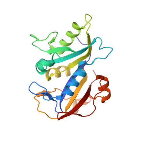
1VJ3 - PubMed Abstract:
The crystal structure of the ternary complex of NADPH, the potent antifolate [2, 4-diamino-5-¿3-[3-(2-acetyloxyethyl)-3-benzyltriazen-1-yl]-4 -chloroph enyl¿-6-ethylpyrimidine] (TAB, 1) and Pneumocystis carinii dihydrofolate reductase (pcDHFR), refined to 2.1 A resolution, reveals that TAB binds similar to the antifolates trimethoprim and methotrexate. These data also reveal multiple conformations for the binding geometry of TAB with two preferred orientations of the acetyloxy and benzyl groups that results from a 180 degrees rotation about the N2-N3 triazenyl bond. The methyl of the acetyloxy and benzyl ring of TAB probes large hydrophobic regions of the p-aminobenzoyl folate binding pocket of the active site, in particular the region near Phe69, which is unique to the pcDHFR sequence. These results confirm prior molecular modeling investigations of the binding of TAB to pcDHFR that identified four low-energy binding geometries, two involving rotations about the terminal N(2)-N(3) triazenyl linkage and two involving atropisomerism about the pivotal pyrimethamine-phenyl bond. The primary differences in the molecular dynamics (MD) models and those observed in this crystal complex result from small conformational changes in active-site residues on energy minimization. However, two MD models place the acetyloxy and benzyl ring groups in a region of the active site between the cofactor-binding region and the p-aminobenzoyl folate pocket; an orientation never observed in any DHFR crystal structure to date. These conformers interact with solvent near the enzyme surface and are probably not observed due to the loss of specific hydrogen bonds with the enzyme. The high species pcDHFR selectivity of TAB could be the result of ligand flexibility that enables multiple binding orientations at the enzyme active site. Further modification of the acetyloxy region of TAB could increase its potency and selectivity for pcDHFR.
Organizational Affiliation:
Hauptman-Woodward Medical Research Institute, Inc., Buffalo, New York 14203, USA. cody@hwi.buffalo.edu