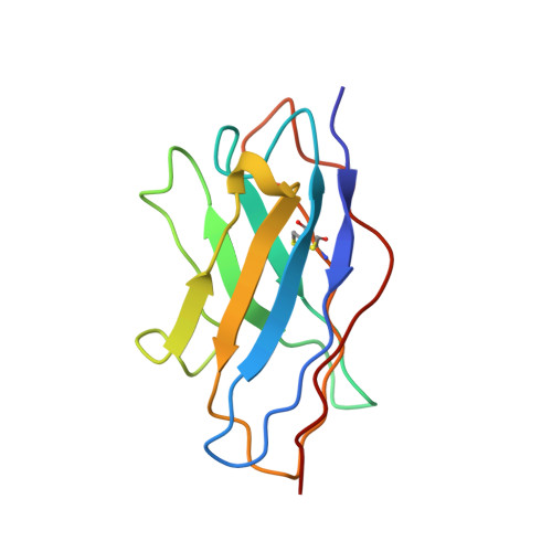
Rearrangement of the former VL interface in the solution structure of a camelised, single antibody VH domain.
Riechmann, L.(1996) J Mol Biol 259: 957-969
- PubMed: 8683598
- DOI: https://doi.org/10.1006/jmbi.1996.0373
- Primary Citation of Related Structures:
1VHP - PubMed Abstract:
The solution structure of the isolated antibody heavy chain variable domain (VH)-P8 was determined by NMR spectroscopy. The VH had previously been modified (camelised) at three positions in its former antibody light chain variable domain (VL) interface to reduce hydrophobicity by mimicking camelid heavy chains naturally devoid of light chains. The architecture of two pleated beta-sheets and the conformation of the H1 and H2 loops in VH-P8 are very similar to those in non-camelised, VL-associated VH domains. Major differences concern the H3 loop, which no longer points towards the now absent VL, and three residues in the former VL interface. The side-chains of Val37 and Trp103 are buried and the Arg38 side-chain exposed in VH-P8. In non-camelised, VL-associated VH domains the side-chains of Val37 and Trp103 are in contact with the VL while the Arg38 side-chain is buried within the VH. Reorientation of Trp103 is due to the local structure in the beta-bulge of strand G. Reorientation of Val37 and Arg38 is caused by a disruption of regular beta-structure in strand C opposite the beta-bulge in strand C'. These changes, combined with the more hydrophilic side-chains of the camelised residues, reduce hydrophobicity and prevent non-specific binding of camelised VH domains, which proved critical for their use as small recognition units. The VH-P8 structure also indicates structural reasons for two other mutations specific for light-chain-lacking camel immunoglobins. Absence of the VH-typical Arg94/Asp101 salt bridge at the base of the H3 loop in VH-P8 may explain why a positively charged residue at position 94 is not conserved in camels. Reorientation of Val37 suggests a function of the camel-specific phenylalanine residue at this position in the hydrophobic core of light-chain-lacking camel heavy chains.
Organizational Affiliation:
MRC Laboratory of Molecular Biology, Cambridge, UK.