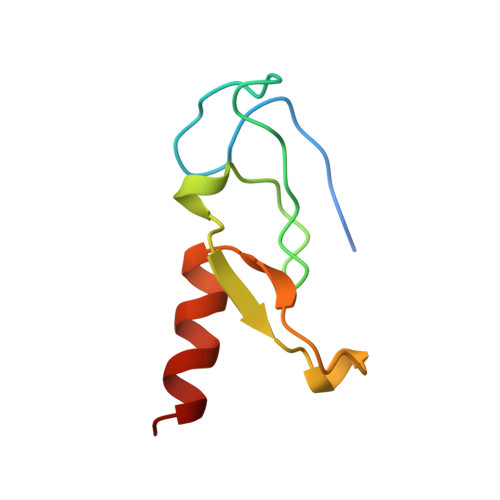
1VFY

PHOSPHATIDYLINOSITOL-3-PHOSPHATE BINDING FYVE DOMAIN OF VPS27P PROTEIN FROM SACCHAROMYCES CEREVISIAE


Experimental Data Snapshot

  • Method: X-RAY DIFFRACTION
  • Resolution: 1.15 Å
  • R-Value Free: 0.181 
  • R-Value Work: 0.174 

wwPDB Validation   3D Report Full Report


This is version 1.4 of the entry. See complete history


Literature

Crystal structure of a phosphatidylinositol 3-phosphate-specific membrane-targeting motif, the FYVE domain of Vps27p.

Misra, S.Hurley, J.H.

(1999) Cell 97: 657-666

  • DOI: https://doi.org/10.1016/s0092-8674(00)80776-x
  • Primary Citation of Related Structures:  
    1VFY

  • PubMed Abstract: 

    Phosphatidylinositol 3-phosphate regulates membrane trafficking and signaling pathways by interacting with the FYVE domains of target proteins. The 1.15 A structure of the Vps27p FYVE domain reveals two antiparallel beta sheets and an alpha helix stabilized by two Zn2+-binding clusters. The core secondary structures are similar to a rabphilin-3A Zn2+-binding domain and to the C1 and LIM domains. Phosphatidylinositol 3-phosphate binds to a pocket formed by the (R/K)(R/K)HHCR motif. A lattice contact shows how anionic ligands can interact with the phosphatidylinositol 3-phosphate-binding site. The tip of the FYVE domain has basic and hydrophobic surfaces positioned so that nonspecific interactions with the phospholipid bilayer can abet specific binding to phosphatidylinositol 3-phosphate.


  • Organizational Affiliation

    Laboratory of Molecular Biology, National Institute of Digestive, Diabetes, and Kidney Diseases, National Institutes of Health, Bethesda, Maryland 20892-0580, USA.


Macromolecules
Find similar proteins by:  (by identity cutoff)  |  3D Structure
Entity ID: 1
MoleculeChains Sequence LengthOrganismDetailsImage
PHOSPHATIDYLINOSITOL-3-PHOSPHATE BINDING FYVE DOMAIN OF PROTEIN VPS2773Saccharomyces cerevisiaeMutation(s): 1 
Gene Names: VPS27
Membrane Entity: Yes 
UniProt
Find proteins for P40343 (Saccharomyces cerevisiae (strain ATCC 204508 / S288c))
Explore P40343 
Go to UniProtKB:  P40343
Entity Groups  
Sequence Clusters30% Identity50% Identity70% Identity90% Identity95% Identity100% Identity
UniProt GroupP40343
Sequence Annotations
Expand
  • Reference Sequence
Experimental Data & Validation

Experimental Data

  • Method: X-RAY DIFFRACTION
  • Resolution: 1.15 Å
  • R-Value Free: 0.181 
  • R-Value Work: 0.174 
  • Space Group: P 1
Unit Cell:
Length ( Å )Angle ( ˚ )
a = 24.09α = 111.79
b = 26.57β = 92.7
c = 31.61γ = 105.7
Software Package:
Software NamePurpose
DENZOdata reduction
SCALEPACKdata scaling
SOLVEphasing
CNSrefinement

Structure Validation

View Full Validation Report


Entry History 

Deposition Data

Revision History  (Full details and data files)

  • Version 1.0: 1999-05-06
    Type: Initial release
  • Version 1.1: 2007-10-16
    Changes: Version format compliance
  • Version 1.2: 2011-07-13
    Changes: Version format compliance
  • Version 1.3: 2021-11-03
    Changes: Database references, Derived calculations
  • Version 1.4: 2023-12-27
    Changes: Data collection