


Crystal Structure of T.th. HB8 Threonine deaminase
Goto, M.To be published.
Experimental Data Snapshot
wwPDB Validation 3D Report Full Report

Entity ID: 1 | |||||
|---|---|---|---|---|---|
| Molecule | Chains | Sequence Length | Organism | Details | Image |
| Threonine deaminase | 311 | Thermus thermophilus HB8 | Mutation(s): 0 EC: 4.3.1.19 | 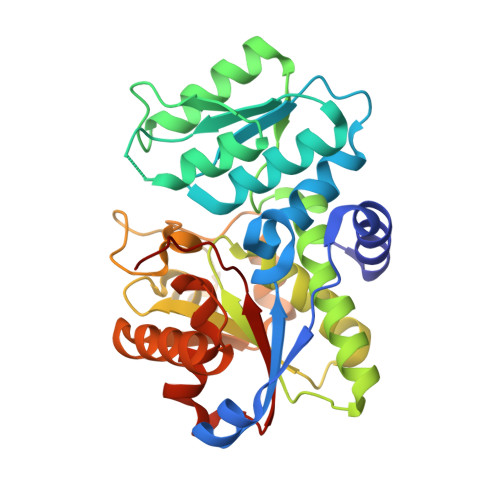 | |
UniProt | |||||
Find proteins for Q5SLL4 (Thermus thermophilus (strain ATCC 27634 / DSM 579 / HB8)) Explore Q5SLL4 Go to UniProtKB: Q5SLL4 | |||||
Entity Groups | |||||
| Sequence Clusters | 30% Identity50% Identity70% Identity90% Identity95% Identity100% Identity | ||||
| UniProt Group | Q5SLL4 | ||||
Sequence AnnotationsExpand | |||||
| |||||
| Ligands 2 Unique | |||||
|---|---|---|---|---|---|
| ID | Chains | Name / Formula / InChI Key | 2D Diagram | 3D Interactions | |
| PLP Query on PLP | F [auth A], H [auth B], J [auth C], L [auth D] | PYRIDOXAL-5'-PHOSPHATE C8 H10 N O6 P NGVDGCNFYWLIFO-UHFFFAOYSA-N |  | ||
| CA Query on CA | E [auth A], G [auth B], I [auth C], K [auth D] | CALCIUM ION Ca BHPQYMZQTOCNFJ-UHFFFAOYSA-N |  | ||
| Length ( Å ) | Angle ( ˚ ) |
|---|---|
| a = 53.59 | α = 90 |
| b = 158.19 | β = 108.52 |
| c = 80.3 | γ = 90 |
| Software Name | Purpose |
|---|---|
| CNS | refinement |
| MOSFLM | data reduction |
| CCP4 | data scaling |
| AMoRE | phasing |
RCSB PDB (citation) is hosted by
RCSB PDB is a member of the

