1VE3

Crystal structure of PH0226 protein from Pyrococcus horikoshii OT3


Experimental Data Snapshot

  • Method: X-RAY DIFFRACTION
  • Resolution: 2.10 Å
  • R-Value Free: 0.270 
  • R-Value Work: 0.236 
  • R-Value Observed: 0.236 

wwPDB Validation   3D Report Full Report


Ligand Structure Quality Assessment 


This is version 1.6 of the entry. See complete history


Literature

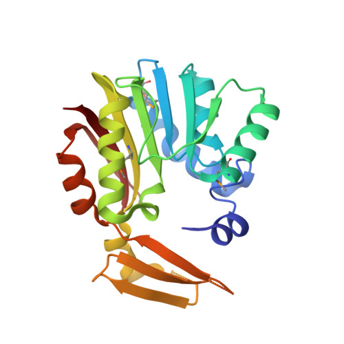
Crystal structure of SAM-dependent methyltransferase from Pyrococcus horikoshii.

Pampa, K.J.Madan Kumar, S.Hema, M.K.Kumara, K.Naveen, S.Kunishima, N.Lokanath, N.K.

(2017) Acta Crystallogr F Struct Biol Commun 73: 706-712

  • DOI: https://doi.org/10.1107/S2053230X17016648
  • Primary Citation of Related Structures:  
    1VE3

  • PubMed Abstract: 

    Methyltransferases (MTs) are enzymes involved in methylation that are needed to perform cellular processes such as biosynthesis, metabolism, gene expression, protein trafficking and signal transduction. The cofactor S-adenosyl-L-methionine (SAM) is used for catalysis by SAM-dependent methyltransferases (SAM-MTs). The crystal structure of Pyrococcus horikoshii SAM-MT was determined to a resolution of 2.1 Å using X-ray diffraction. The monomeric structure consists of a Rossmann-like fold (domain I) and a substrate-binding domain (domain II). The cofactor (SAM) molecule binds at the interface between adjacent subunits, presumably near to the active site(s) of the enzyme. The observed dimeric state might be important for the catalytic function of the enzyme.


  • Organizational Affiliation

    Department of Studies in Biotechnology, University of Mysore, Manasagangotri, Mysuru, Karnataka 570 006, India.


Macromolecules
Find similar proteins by:  (by identity cutoff)  |  3D Structure
Entity ID: 1
MoleculeChains Sequence LengthOrganismDetailsImage
hypothetical protein PH0226
A, B
227Pyrococcus horikoshiiMutation(s): 0 
UniProt
Find proteins for O57965 (Pyrococcus horikoshii (strain ATCC 700860 / DSM 12428 / JCM 9974 / NBRC 100139 / OT-3))
Explore O57965 
Go to UniProtKB:  O57965
Entity Groups  
Sequence Clusters30% Identity50% Identity70% Identity90% Identity95% Identity100% Identity
UniProt GroupO57965
Sequence Annotations
Expand
  • Reference Sequence
Small Molecules
Modified Residues  1 Unique
IDChains TypeFormula2D DiagramParent
MSE
Query on MSE
A, B
L-PEPTIDE LINKINGC5 H11 N O2 SeMET
Experimental Data & Validation

Experimental Data

  • Method: X-RAY DIFFRACTION
  • Resolution: 2.10 Å
  • R-Value Free: 0.270 
  • R-Value Work: 0.236 
  • R-Value Observed: 0.236 
  • Space Group: P 32 2 1
Unit Cell:
Length ( Å )Angle ( ˚ )
a = 58.167α = 90
b = 58.167β = 90
c = 252.463γ = 120
Software Package:
Software NamePurpose
CNSrefinement
HKL-2000data reduction
SCALEPACKdata scaling
SOLVEphasing

Structure Validation

View Full Validation Report


Ligand Structure Quality Assessment 


Entry History 

Deposition Data

Revision History  (Full details and data files)

  • Version 1.0: 2005-05-24
    Type: Initial release
  • Version 1.1: 2008-04-27
    Changes: Version format compliance
  • Version 1.2: 2011-07-13
    Changes: Version format compliance
  • Version 1.3: 2017-12-13
    Changes: Database references
  • Version 1.4: 2020-01-01
    Changes: Database references, Derived calculations
  • Version 1.5: 2020-01-22
    Changes: Derived calculations
  • Version 1.6: 2023-12-27
    Changes: Data collection, Database references, Derived calculations