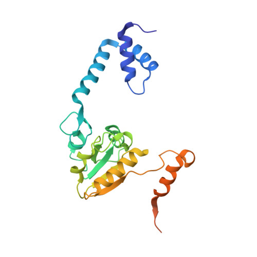
1VDD

Crystal structure of recombinational repair protein RecR


Experimental Data Snapshot

  • Method: X-RAY DIFFRACTION
  • Resolution: 2.50 Å
  • R-Value Free: 0.301 
  • R-Value Work: 0.233 
  • R-Value Observed: 0.233 

wwPDB Validation   3D Report Full Report


This is version 1.4 of the entry. See complete history


Literature

Ring-shaped architecture of RecR: implications for its role in homologous recombinational DNA repair

Lee, B.I.Kim, K.H.Park, S.J.Eom, S.H.Song, H.K.Suh, S.W.

(2004) EMBO J 23: 2029-2038

  • DOI: https://doi.org/10.1038/sj.emboj.7600222
  • Primary Citation of Related Structures:  
    1VDD

  • PubMed Abstract: 

    RecR, together with RecF and RecO, facilitates RecA loading in the RecF pathway of homologous recombinational DNA repair in procaryotes. The human Rad52 protein is a functional counterpart of RecFOR. We present here the crystal structure of RecR from Deinococcus radiodurans (DR RecR). A monomer of DR RecR has a two-domain structure: the N-terminal domain with a helix-hairpin-helix (HhH) motif and the C-terminal domain with a Cys4 zinc-finger motif, a Toprim domain and a Walker B motif. Four such monomers form a ring-shaped tetramer of 222 symmetry with a central hole of 30-35 angstroms diameter. In the crystal, two tetramers are concatenated, implying that the RecR tetramer is capable of opening and closing. We also show that DR RecR binds to both dsDNA and ssDNA, and that its HhH motif is essential for DNA binding.


  • Organizational Affiliation

    Department of Chemistry, College of Natural Sciences, Seoul National University, Seoul, Korea.


Macromolecules
Find similar proteins by:  (by identity cutoff)  |  3D Structure
Entity ID: 1
MoleculeChains Sequence LengthOrganismDetailsImage
Recombination protein recR
A, B, C, D
228Deinococcus radioduransMutation(s): 0 
Gene Names: recR
UniProt
Find proteins for Q9ZNA2 (Deinococcus radiodurans (strain ATCC 13939 / DSM 20539 / JCM 16871 / CCUG 27074 / LMG 4051 / NBRC 15346 / NCIMB 9279 / VKM B-1422 / R1))
Explore Q9ZNA2 
Go to UniProtKB:  Q9ZNA2
Entity Groups  
Sequence Clusters30% Identity50% Identity70% Identity90% Identity95% Identity100% Identity
UniProt GroupQ9ZNA2
Sequence Annotations
Expand
  • Reference Sequence
Experimental Data & Validation

Experimental Data

  • Method: X-RAY DIFFRACTION
  • Resolution: 2.50 Å
  • R-Value Free: 0.301 
  • R-Value Work: 0.233 
  • R-Value Observed: 0.233 
  • Space Group: C 2 2 21
Unit Cell:
Length ( Å )Angle ( ˚ )
a = 106.218α = 90
b = 121.386β = 90
c = 154.993γ = 90
Software Package:
Software NamePurpose
CNSrefinement
SMARTdata reduction
SAINTPLUSdata scaling
LSCALEdata scaling
SOLVEphasing

Structure Validation

View Full Validation Report


Entry History 

Deposition Data

Revision History  (Full details and data files)

  • Version 1.0: 2004-05-18
    Type: Initial release
  • Version 1.1: 2008-04-27
    Changes: Version format compliance
  • Version 1.2: 2011-07-13
    Changes: Version format compliance
  • Version 1.3: 2017-10-04
    Changes: Refinement description
  • Version 1.4: 2023-12-27
    Changes: Data collection, Database references, Derived calculations