1V9D

Crystal structure of the core FH2 domain of mouse mDia1


Experimental Data Snapshot

  • Method: X-RAY DIFFRACTION
  • Resolution: 2.60 Å
  • R-Value Free: 0.267 
  • R-Value Work: 0.236 
  • R-Value Observed: 0.239 

wwPDB Validation   3D Report Full Report


This is version 1.3 of the entry. See complete history


Literature

The core FH2 domain of diaphanous-related formins is an elongated actin binding protein that inhibits polymerization.

Shimada, A.Nyitrai, M.Vetter, I.R.Kuhlmann, D.Bugyi, B.Narumiya, S.Geeves, M.A.Wittinghofer, A.

(2004) Mol Cell 13: 511-522

  • DOI: https://doi.org/10.1016/s1097-2765(04)00059-0
  • Primary Citation of Related Structures:  
    1V9D

  • PubMed Abstract: 

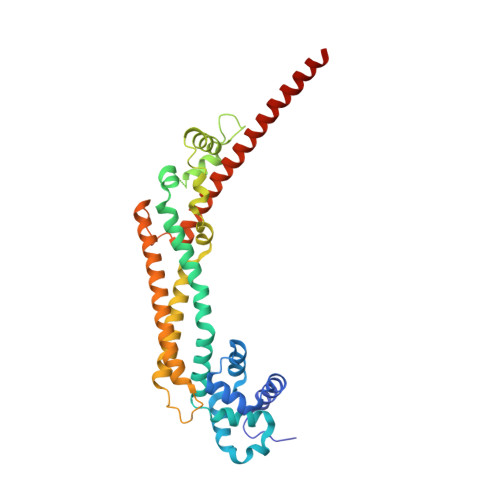
    Diaphanous-related formins (Drf) are activated by Rho GTP binding proteins and induce polymerization of unbranched actin filaments. They contain three formin homology domains. Evidence as to the effect of formins on actin polymerization were obtained using FH2/FH1 constructs of various length from different Drfs. Here we define the core FH2 domain as a proteolytically stable domain of approximately 338 residues. The monomeric FH2 domains from mDia1 and mDia3 inhibit polymerization of actin and can bind in a 1:1 complex with F-actin at micromolar concentrations. The X-ray structure analysis of the domain shows an elongated, crescent-shaped molecule consisting of three helical subdomains. The most highly conserved regions of the domain span a distance of 75 A and are both required for barbed-end inhibition. A construct containing an additional 72 residue linker has dramatically different properties: It oligomerizes and induces actin polymerization at subnanomolar concentration.


  • Organizational Affiliation

    Max-Planck Institut für Molekulare Physiologie, Otto Hahn Strasse 11, D-44227 Dortmund, Germany.


Macromolecules
Find similar proteins by:  (by identity cutoff)  |  3D Structure
Entity ID: 1
MoleculeChains Sequence LengthOrganismDetailsImage
Diaphanous protein homolog 1
A, B, C, D
340Mus musculusMutation(s): 0 
UniProt & NIH Common Fund Data Resources
Find proteins for O08808 (Mus musculus)
Explore O08808 
Go to UniProtKB:  O08808
IMPC:  MGI:1194490
Entity Groups  
Sequence Clusters30% Identity50% Identity70% Identity90% Identity95% Identity100% Identity
UniProt GroupO08808
Sequence Annotations
Expand
  • Reference Sequence
Experimental Data & Validation

Experimental Data

  • Method: X-RAY DIFFRACTION
  • Resolution: 2.60 Å
  • R-Value Free: 0.267 
  • R-Value Work: 0.236 
  • R-Value Observed: 0.239 
  • Space Group: P 21 21 21
Unit Cell:
Length ( Å )Angle ( ˚ )
a = 82.43α = 90
b = 124.521β = 90
c = 229.404γ = 90
Software Package:
Software NamePurpose
HKL-2000data collection
SCALEPACKdata scaling
SHARPphasing
CNSrefinement
HKL-2000data reduction

Structure Validation

View Full Validation Report


Entry History 

Deposition Data

Revision History  (Full details and data files)

  • Version 1.0: 2004-03-09
    Type: Initial release
  • Version 1.1: 2008-04-27
    Changes: Version format compliance
  • Version 1.2: 2011-07-13
    Changes: Version format compliance
  • Version 1.3: 2023-12-27
    Changes: Data collection, Database references, Derived calculations