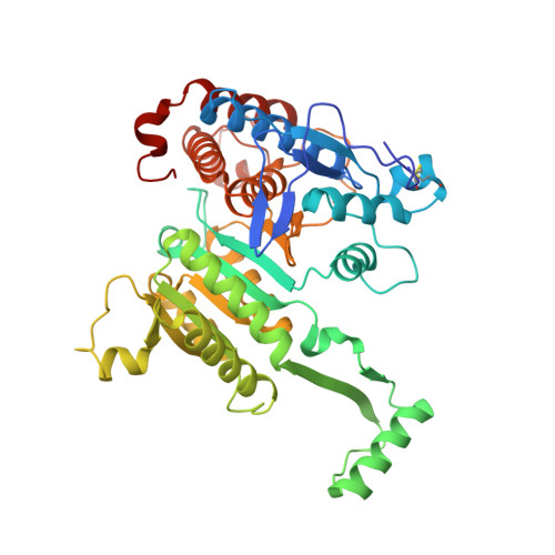
Crystal structure of isocitrate dehydrogenase from Aeropyrum pernix
Jeong, J.-J., Sonoda, T., Fushinobu, S., Shoun, H., Wakagi, T.(2004) Proteins 55: 1087-1089
- PubMed: 15146507
- DOI: https://doi.org/10.1002/prot.20121
- Primary Citation of Related Structures:
1V94
Organizational Affiliation:
Department of Biotechnology, University of Tokyo, 1-1-1 Yayoi, Bunkyo-Ku, Tokyo 113-8657, Japan.