1V74

Structure of the E. coli colicin D bound to its immunity protein ImmD


Experimental Data Snapshot

  • Method: X-RAY DIFFRACTION
  • Resolution: 2.00 Å
  • R-Value Free: 0.234 
  • R-Value Work: 0.194 

wwPDB Validation   3D Report Full Report


This is version 1.4 of the entry. See complete history


Literature

Structural inhibition of the colicin D tRNase by the tRNA-mimicking immunity protein

Graille, M.Mora, L.Buckingham, R.H.Van Tilbeurgh, H.De Zamaroczy, M.

(2004) EMBO J 23: 1474-1482

  • DOI: https://doi.org/10.1038/sj.emboj.7600162
  • Primary Citation of Related Structures:  
    1V74

  • PubMed Abstract: 

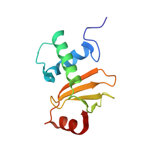
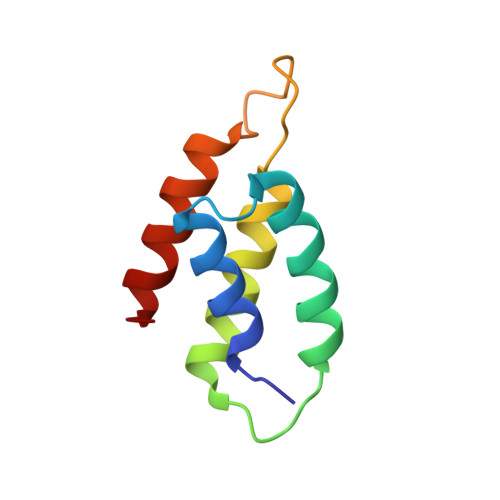
    Colicins are toxins secreted by Escherichia coli in order to kill their competitors. Colicin D is a 75 kDa protein that consists of a translocation domain, a receptor-binding domain and a cytotoxic domain, which specifically cleaves the anticodon loop of all four tRNA(Arg) isoacceptors, thereby inactivating protein synthesis and leading to cell death. Here we report the 2.0 A resolution crystal structure of the complex between the toxic domain and its immunity protein ImmD. Neither component shows structural homology to known RNases or their inhibitors. In contrast to other characterized colicin nuclease-Imm complexes, the colicin D active site pocket is completely blocked by ImmD, which, by bringing a negatively charged cluster in opposition to a positively charged cluster on the surface of colicin D, appears to mimic the tRNA substrate backbone. Site-directed mutations affecting either the catalytic domain or the ImmD protein have led to the identification of the residues vital for catalytic activity and for the tight colicin D/ImmD interaction that inhibits colicin D toxicity and tRNase catalytic activity.


  • Organizational Affiliation

    LEBS, CNRS, UPR 9063, Gif sur Yvette, France.


Macromolecules
Find similar proteins by:  (by identity cutoff)  |  3D Structure
Entity ID: 1
MoleculeChains Sequence LengthOrganismDetailsImage
Colicin D107Escherichia coliMutation(s): 0 
UniProt
Find proteins for P17998 (Escherichia coli)
Explore P17998 
Go to UniProtKB:  P17998
Entity Groups  
Sequence Clusters30% Identity50% Identity70% Identity90% Identity95% Identity100% Identity
UniProt GroupP17998
Sequence Annotations
Expand
  • Reference Sequence
Find similar proteins by:  (by identity cutoff)  |  3D Structure
Entity ID: 2
MoleculeChains Sequence LengthOrganismDetailsImage
Colicin D immunity protein87Escherichia coliMutation(s): 0 
UniProt
Find proteins for P11899 (Escherichia coli)
Explore P11899 
Go to UniProtKB:  P11899
Entity Groups  
Sequence Clusters30% Identity50% Identity70% Identity90% Identity95% Identity100% Identity
UniProt GroupP11899
Sequence Annotations
Expand
  • Reference Sequence
Small Molecules
Ligands 1 Unique
IDChains Name / Formula / InChI Key2D Diagram3D Interactions
1PE
Query on 1PE

Download Ideal Coordinates CCD File 
C [auth A]PENTAETHYLENE GLYCOL
C10 H22 O6
JLFNLZLINWHATN-UHFFFAOYSA-N
Experimental Data & Validation

Experimental Data

  • Method: X-RAY DIFFRACTION
  • Resolution: 2.00 Å
  • R-Value Free: 0.234 
  • R-Value Work: 0.194 
  • Space Group: P 41 21 2
Unit Cell:
Length ( Å )Angle ( ˚ )
a = 62.057α = 90
b = 62.057β = 90
c = 147.934γ = 90
Software Package:
Software NamePurpose
DENZOdata reduction
SCALEPACKdata scaling
SOLVEphasing
CNSrefinement

Structure Validation

View Full Validation Report


Entry History 

Deposition Data

Revision History  (Full details and data files)

  • Version 1.0: 2004-03-30
    Type: Initial release
  • Version 1.1: 2008-04-27
    Changes: Version format compliance
  • Version 1.2: 2011-07-13
    Changes: Version format compliance
  • Version 1.3: 2018-01-03
    Changes: Structure summary
  • Version 1.4: 2023-12-27
    Changes: Data collection, Database references, Derived calculations