


The crystal structure of 3-isopropylmalate dehydrogenase from Bacillus coagulans
Fujita, K., Minami, H., Suzuki, K., Tsunoda, M., Sekiguchi, T., Mizui, R., Tsuzaki, S., Nakamura, S., Takenaka, A.To be published.
Experimental Data Snapshot
wwPDB Validation 3D Report Full Report

Entity ID: 1 | |||||
|---|---|---|---|---|---|
| Molecule | Chains | Sequence Length | Organism | Details | Image |
| 3-isopropylmalate dehydrogenase | 366 | Heyndrickxia coagulans | Mutation(s): 0 EC: 1.1.1.85 | 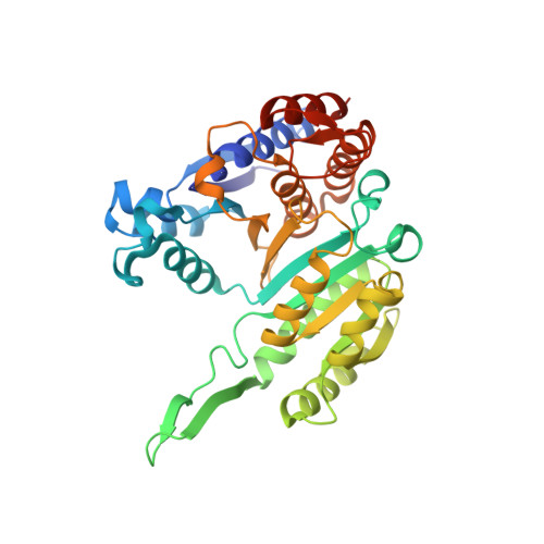 | |
UniProt | |||||
Find proteins for P12010 (Heyndrickxia coagulans) Explore P12010 Go to UniProtKB: P12010 | |||||
Entity Groups | |||||
| Sequence Clusters | 30% Identity50% Identity70% Identity90% Identity95% Identity100% Identity | ||||
| UniProt Group | P12010 | ||||
Sequence AnnotationsExpand | |||||
| |||||
| Length ( Å ) | Angle ( ˚ ) |
|---|---|
| a = 112.18 | α = 90 |
| b = 112.18 | β = 90 |
| c = 192.07 | γ = 120 |
| Software Name | Purpose |
|---|---|
| d*TREK | data scaling |
| SCALA | data scaling |
| AMoRE | phasing |
| CNS | refinement |
| d*TREK | data reduction |
| CCP4 | data scaling |
RCSB PDB (citation) is hosted by
RCSB PDB is a member of the

