1V4S

Crystal structure of human glucokinase


Experimental Data Snapshot

  • Method: X-RAY DIFFRACTION
  • Resolution: 2.30 Å
  • R-Value Free: 0.273 
  • R-Value Work: 0.232 
  • R-Value Observed: 0.235 

wwPDB Validation   3D Report Full Report


Ligand Structure Quality Assessment 


This is version 1.5 of the entry. See complete history


Literature

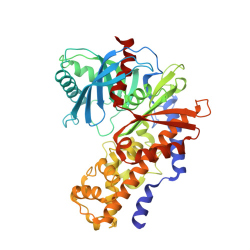
Structural basis for allosteric regulation of the monomeric allosteric enzyme human glucokinase

Kamata, K.Mitsuya, M.Nishimura, T.Eiki, J.Nagata, Y.

(2004) Structure 12: 429-438

  • DOI: https://doi.org/10.1016/j.str.2004.02.005
  • Primary Citation of Related Structures:  
    1V4S, 1V4T

  • PubMed Abstract: 

    Glucokinase is a monomeric enzyme that displays a low affinity for glucose and a sigmoidal saturation curve for its substrate, two properties that are important for its playing the role of a glucose sensor in pancreas and liver. The molecular basis for these two properties is not well understood. Herein we report the crystal structures of glucokinase in its active and inactive forms, which demonstrate that global conformational change, including domain reorganization, is induced by glucose binding. This suggests that the positive cooperativity of monomeric glucokinase obeys the "mnemonical mechanism" rather than the well-known concerted model. These structures also revealed an allosteric site through which small molecules may modulate the kinetic properties of the enzyme. This finding provided the mechanistic basis for activation of glucokinase as a potential therapeutic approach for treating type 2 diabetes mellitus.


  • Organizational Affiliation

    Banyu Tsukuba Research Institute in collaboration with Merck Research Laboratories, Okubo 3, Tsukuba, Ibaraki 300-2611, Japan. kenji_kamata@merck.com


Macromolecules
Find similar proteins by:  (by identity cutoff)  |  3D Structure
Entity ID: 1
MoleculeChains Sequence LengthOrganismDetailsImage
glucokinase isoform 2455Homo sapiensMutation(s): 0 
EC: 2.7.1.1
UniProt & NIH Common Fund Data Resources
Find proteins for P35557 (Homo sapiens)
Explore P35557 
Go to UniProtKB:  P35557
PHAROS:  P35557
GTEx:  ENSG00000106633 
Entity Groups  
Sequence Clusters30% Identity50% Identity70% Identity90% Identity95% Identity100% Identity
UniProt GroupP35557
Sequence Annotations
Expand
  • Reference Sequence
Small Molecules
Ligands 3 Unique
IDChains Name / Formula / InChI Key2D Diagram3D Interactions
MRK
Query on MRK

Download Ideal Coordinates CCD File 
D [auth A]2-AMINO-4-FLUORO-5-[(1-METHYL-1H-IMIDAZOL-2-YL)SULFANYL]-N-(1,3-THIAZOL-2-YL)BENZAMIDE
C14 H12 F N5 O S2
YUCYMQBDBXVNCE-UHFFFAOYSA-N
GLC
Query on GLC

Download Ideal Coordinates CCD File 
B [auth A]alpha-D-glucopyranose
C6 H12 O6
WQZGKKKJIJFFOK-DVKNGEFBSA-N
NA
Query on NA

Download Ideal Coordinates CCD File 
C [auth A]SODIUM ION
Na
FKNQFGJONOIPTF-UHFFFAOYSA-N
Binding Affinity Annotations 
IDSourceBinding Affinity
MRK BindingDB:  1V4S Kd: 360 (nM) from 1 assay(s)
EC50: 67.11 (nM) from 1 assay(s)
Experimental Data & Validation

Experimental Data

  • Method: X-RAY DIFFRACTION
  • Resolution: 2.30 Å
  • R-Value Free: 0.273 
  • R-Value Work: 0.232 
  • R-Value Observed: 0.235 
  • Space Group: P 65 2 2
Unit Cell:
Length ( Å )Angle ( ˚ )
a = 79.855α = 90
b = 79.855β = 90
c = 322.221γ = 120
Software Package:
Software NamePurpose
CNXrefinement
DENZOdata reduction
SCALEPACKdata scaling
AMoREphasing

Structure Validation

View Full Validation Report


Ligand Structure Quality Assessment 


Entry History 

Deposition Data

Revision History  (Full details and data files)

  • Version 1.0: 2004-03-30
    Type: Initial release
  • Version 1.1: 2008-04-27
    Changes: Version format compliance
  • Version 1.2: 2011-07-13
    Changes: Version format compliance
  • Version 1.3: 2020-07-29
    Type: Remediation
    Reason: Carbohydrate remediation
    Changes: Data collection, Derived calculations, Structure summary
  • Version 1.4: 2022-12-21
    Changes: Database references, Structure summary
  • Version 1.5: 2023-11-08
    Changes: Data collection, Refinement description