Understanding protein-ligand interactions: the price of protein flexibility
Rauh, D., Klebe, G., Stubbs, M.T.(2004) J Mol Biol 335: 1325-1341
- PubMed: 14729347
- DOI: https://doi.org/10.1016/j.jmb.2003.11.041
- Primary Citation of Related Structures:
1V2J, 1V2K, 1V2L, 1V2M, 1V2N, 1V2O, 1V2P, 1V2Q, 1V2R, 1V2S, 1V2T, 1V2U, 1V2V, 1V2W - PubMed Abstract:
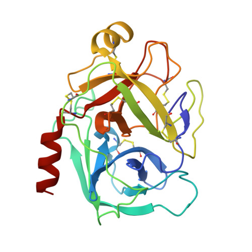
In order to design selective, high-affinity ligands to a target protein, it is advantageous to understand the structural determinants for protein-ligand complex formation at the atomic level. In a model system, we have successively mapped the factor Xa binding site onto trypsin, showing that certain mutations influence both protein structure and inhibitor specificity. Our previous studies have shown that introduction of the 172SSFI175 sequence of factor Xa into rat or bovine trypsin results in the destabilisation of the intermediate helix with burial of Phe174 (the down conformation). Surface exposure of the latter residue (the up conformation) is critical for the correct formation of the aromatic box found in factor Xa-ligand complexes. In the present study, we investigate the influence of aromatic residues in position 174. Replacement with the bulky tryptophan (SSWI) shows reduced affinity for benzamidine-based inhibitors (1) and (4), whereas removal of the side-chain (alanine, SSAI) or exchange with a hydrophilic residue (arginine, SSRI) leads to a significant loss in affinity for all inhibitors studied. The variants could be crystallised in the presence of different inhibitors in multiple crystal forms. Structural characterisation of the variants revealed three different conformations of the intermediate helix and 175 loop in SSAI (down, up and super-up), as well as a complete disorder of this region in one crystal form of SSRI, suggesting that the compromised affinity of these variants is related to conformational flexibility. The influence of Glu217, peripheral to the ligand-binding site in factor Xa, was investigated. Introduction of Glu217 into trypsin variants containing the SSFI sequence exhibited enhanced affinity for the factor Xa ligands (2) and (3). The crystal structures of these variants also exhibited the down and super-up conformations, the latter of which could be converted to up upon soaking and binding of inhibitor (2). The improved affinity of the Glu217-containing variants appears to be due to a shift towards the up conformation. Thus, the reduction in affinity caused by conformational variability of the protein target can be partially or wholly offset by compensatory binding to the up conformation. The insights provided by these studies will be helpful in improving our understanding of ligand binding for the drug design process.
Organizational Affiliation:
Institut für Pharmazeutische Chemie der Philipps, Universität Marburg, Marbacher Weg 6, D35032 Marburg, Germany.