1V2H

Crystal structure of human PNP complexed with guanine


Experimental Data Snapshot

  • Method: X-RAY DIFFRACTION
  • Resolution: 2.70 Å
  • R-Value Free: 0.293 
  • R-Value Work: 0.218 
  • R-Value Observed: 0.218 

wwPDB Validation   3D Report Full Report


Ligand Structure Quality Assessment 


This is version 1.3 of the entry. See complete history


Literature

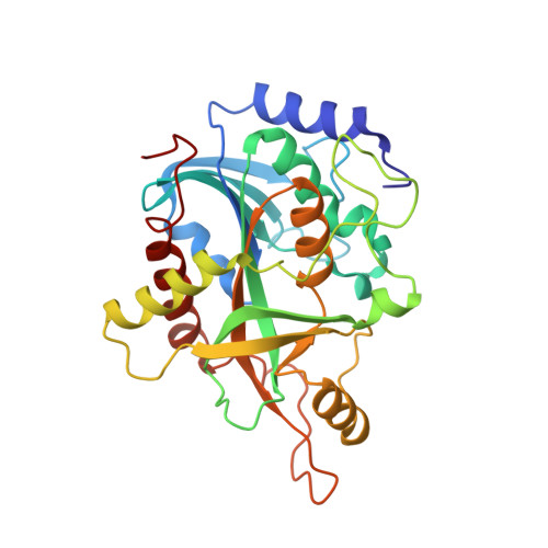
Crystal structure of human PNP complexed with guanine.

de Azevedo Jr., W.F.Canduri, F.dos Santos, D.M.Pereira, J.H.Bertacine Dias, M.V.Silva, R.G.Mendes, M.A.Basso, L.A.Palma, M.S.Santos, D.S.

(2003) Biochem Biophys Res Commun 312: 767-772

  • DOI: https://doi.org/10.1016/j.bbrc.2003.10.190
  • Primary Citation of Related Structures:  
    1V2H

  • PubMed Abstract: 

    Purine nucleoside phosphorylase (PNP) catalyzes the phosphorolysis of the N-ribosidic bonds of purine nucleosides and deoxynucleosides. PNP is a target for inhibitor development aiming at T-cell immune response modulation and has been submitted to extensive structure-based drug design. More recently, the 3-D structure of human PNP has been refined to 2.3A resolution, which allowed a redefinition of the residues involved in the substrate-binding sites and provided a more reliable model for structure-based design of inhibitors. This work reports crystallographic study of the complex of Human PNP:guanine (HsPNP:Gua) solved at 2.7A resolution using synchrotron radiation. Analysis of the structural differences among the HsPNP:Gua complex, PNP apoenzyme, and HsPNP:immucillin-H provides explanation for inhibitor binding, refines the purine-binding site, and can be used for future inhibitor design.


  • Organizational Affiliation

    Departamento de Física, UNESP, São José do Rio Preto, SP 15054-000, Brazil. walterfa@df.ibilce.unesp.br


Macromolecules
Find similar proteins by:  (by identity cutoff)  |  3D Structure
Entity ID: 1
MoleculeChains Sequence LengthOrganismDetailsImage
Purine nucleoside phosphorylaseA [auth E]288Homo sapiensMutation(s): 0 
Gene Names: PNP
EC: 2.4.2.1
UniProt & NIH Common Fund Data Resources
Find proteins for P00491 (Homo sapiens)
Explore P00491 
Go to UniProtKB:  P00491
PHAROS:  P00491
GTEx:  ENSG00000198805 
Entity Groups  
Sequence Clusters30% Identity50% Identity70% Identity90% Identity95% Identity100% Identity
UniProt GroupP00491
Sequence Annotations
Expand
  • Reference Sequence
Experimental Data & Validation

Experimental Data

  • Method: X-RAY DIFFRACTION
  • Resolution: 2.70 Å
  • R-Value Free: 0.293 
  • R-Value Work: 0.218 
  • R-Value Observed: 0.218 
  • Space Group: H 3 2
Unit Cell:
Length ( Å )Angle ( ˚ )
a = 141.07α = 90
b = 141.07β = 90
c = 162.37γ = 120
Software Package:
Software NamePurpose
MOSFLMdata reduction
SCALAdata scaling
AMoREphasing
X-PLORrefinement
CCP4data scaling

Structure Validation

View Full Validation Report


Ligand Structure Quality Assessment 


Entry History 

Revision History  (Full details and data files)

  • Version 1.0: 2004-01-13
    Type: Initial release
  • Version 1.1: 2008-04-27
    Changes: Version format compliance
  • Version 1.2: 2011-07-13
    Changes: Derived calculations, Version format compliance
  • Version 1.3: 2023-10-25
    Changes: Data collection, Database references, Derived calculations, Refinement description