1V00

Erythrina cristagalli lectin


Experimental Data Snapshot

  • Method: X-RAY DIFFRACTION
  • Resolution: 1.70 Å
  • R-Value Free: 0.195 
  • R-Value Work: 0.172 
  • R-Value Observed: 0.172 

wwPDB Validation   3D Report Full Report


This is version 2.0 of the entry. See complete history


Literature

Crystal Structures of Erythrina Cristagalli Lectin with Bound N-Linked Oligosaccharide and Lactose

Turton, K.Natesh, R.Thiyagarajan, N.Chaddock, J.A.Acharya, K.R.

(2004) Glycobiology 14: 923

  • DOI: https://doi.org/10.1093/glycob/cwh114
  • Primary Citation of Related Structures:  
    1UZY, 1UZZ, 1V00

  • PubMed Abstract: 

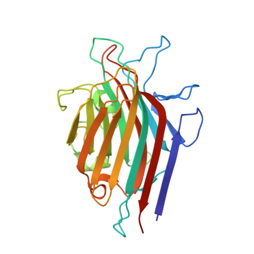
    Erythrina cristagalli lectin (ECL) is a galactose-specific legume lectin. Although its biological function in the legume is unknown, ECL exhibits hemagglutinating activity in vitro and is mitogenic for T lymphocytes. In addition, it has been recently shown that ECL forms a novel conjugate when coupled to a catalytically active derivative of the type A neurotoxin from Clostridium botulinum, thus providing a therapeutic potential. ECL is biologically active as a dimer in which each protomer contains a functional carbohydrate-combining site. The crystal structure of native ECL was recently reported in complex with lactose and 2'-fucosyllactose. ECL protomers adopt the legume lectin fold but form non-canonical dimers via the handshake motif as was previously observed for Erythrina corallodendron lectin. Here we report the crystal structures of native and recombinant forms of the lectin in three new crystal forms, both unliganded and in complex with lactose. For the first time, the detailed structure of the glycosylated hexasaccharide for native ECL has been elucidated. The structure also shows that in the crystal lattice the glycosylation site and the carbohydrate binding site are involved in intermolecular contacts through water-mediated interactions.


  • Organizational Affiliation

    Department of Biology and Biochemistry, University of Bath, Claverton Down, Bath BA2 7AY, United Kingdom.


Macromolecules
Find similar proteins by:  (by identity cutoff)  |  3D Structure
Entity ID: 1
MoleculeChains Sequence LengthOrganismDetailsImage
LECTIN (ECL)
A, B, C, D
242Erythrina crista-galliMutation(s): 0 
UniProt
Find proteins for Q6YD91 (Erythrina crista-galli)
Explore Q6YD91 
Go to UniProtKB:  Q6YD91
Entity Groups  
Sequence Clusters30% Identity50% Identity70% Identity90% Identity95% Identity100% Identity
UniProt GroupQ6YD91
Sequence Annotations
Expand
  • Reference Sequence
Oligosaccharides

Help

Entity ID: 2
MoleculeChains Length2D Diagram Glycosylation3D Interactions
beta-D-galactopyranose-(1-4)-beta-D-glucopyranose
E, F, G, H
2N/A
Glycosylation Resources
GlyTouCan:  G84224TW
GlyCosmos:  G84224TW
GlyGen:  G84224TW
Biologically Interesting Molecules (External Reference) 1 Unique
Experimental Data & Validation

Experimental Data

  • Method: X-RAY DIFFRACTION
  • Resolution: 1.70 Å
  • R-Value Free: 0.195 
  • R-Value Work: 0.172 
  • R-Value Observed: 0.172 
  • Space Group: P 1 21 1
Unit Cell:
Length ( Å )Angle ( ˚ )
a = 54.9α = 90
b = 167.23β = 97.08
c = 55.13γ = 90
Software Package:
Software NamePurpose
CNSrefinement
DENZOdata reduction
SCALEPACKdata scaling
AMoREphasing

Structure Validation

View Full Validation Report


Entry History 

Deposition Data

Revision History  (Full details and data files)

  • Version 1.0: 2004-06-22
    Type: Initial release
  • Version 1.1: 2011-05-07
    Changes: Version format compliance
  • Version 1.2: 2011-07-13
    Changes: Version format compliance
  • Version 2.0: 2020-07-29
    Type: Remediation
    Reason: Carbohydrate remediation
    Changes: Advisory, Atomic model, Data collection, Derived calculations, Non-polymer description, Structure summary