1UZR

Crystal Structure of the Class Ib Ribonucleotide Reductase R2F-2 subunit from Mycobacterium tuberculosis


Experimental Data Snapshot

  • Method: X-RAY DIFFRACTION
  • Resolution: 2.20 Å
  • R-Value Free: 0.202 
  • R-Value Work: 0.175 
  • R-Value Observed: 0.177 

wwPDB Validation   3D Report Full Report


This is version 1.3 of the entry. See complete history


Literature

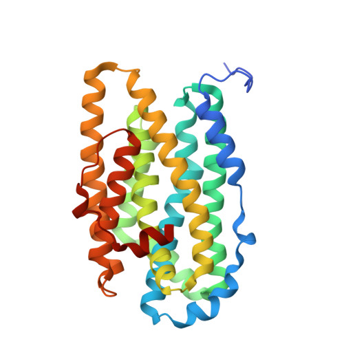
Crystal Structure of the Biologically Active Form of Class Ib Ribonucleotide Reductase Small Subunit from Mycobacterium Tuberculosis

Uppsten, M.Davis, J.Rubin, H.Uhlin, U.

(2004) FEBS Lett 569: 117

  • DOI: https://doi.org/10.1016/j.febslet.2004.05.059
  • Primary Citation of Related Structures:  
    1UZR

  • PubMed Abstract: 

    Two nrdF genes of Mycobacterium tuberculosis code for different R2 subunits of the class Ib ribonucleotide reductase (RNR). The proteins are denoted R2F-1 and R2F-2 having 71% sequence identity. The R2F-2 subunit forms the biologically active RNR complex with the catalytic R1E-subunit. We present the structure of the reduced R2F-2 subunit to 2.2 A resolution. Comparison of the R2F-2 structure with a model of R2F-1 suggests that the important differences are located at the C-terminus. We found that within class Ib, the E-helix close to the iron diiron centre has two preferred conformations, which cannot be explained by the redox-state of the diiron centre. In the R2F-2 structure, we also could see a mobility of alphaE in between the two conformations.


  • Organizational Affiliation

    Department of Molecular Biology, Swedish University of Agricultural Sciences, Uppsala Biomedical Center, P.O. Box 590, SE-751 24 Uppsala, Sweden.


Macromolecules
Find similar proteins by:  (by identity cutoff)  |  3D Structure
Entity ID: 1
MoleculeChains Sequence LengthOrganismDetailsImage
RIBONUCLEOTIDE REDUCTASE R2-2 SMALL SUBUNIT
A, B, C
296Mycobacterium tuberculosisMutation(s): 0 
EC: 1.17.4.1
UniProt
Find proteins for P9WH71 (Mycobacterium tuberculosis (strain ATCC 25618 / H37Rv))
Explore P9WH71 
Go to UniProtKB:  P9WH71
Entity Groups  
Sequence Clusters30% Identity50% Identity70% Identity90% Identity95% Identity100% Identity
UniProt GroupP9WH71
Sequence Annotations
Expand
  • Reference Sequence
Small Molecules
Ligands 3 Unique
IDChains Name / Formula / InChI Key2D Diagram3D Interactions
CIT
Query on CIT

Download Ideal Coordinates CCD File 
F [auth A],
J [auth B],
N [auth C]
CITRIC ACID
C6 H8 O7
KRKNYBCHXYNGOX-UHFFFAOYSA-N
GOL
Query on GOL

Download Ideal Coordinates CCD File 
G [auth A],
K [auth B],
O [auth C]
GLYCEROL
C3 H8 O3
PEDCQBHIVMGVHV-UHFFFAOYSA-N
FE
Query on FE

Download Ideal Coordinates CCD File 
D [auth A]
E [auth A]
H [auth B]
I [auth B]
L [auth C]
D [auth A],
E [auth A],
H [auth B],
I [auth B],
L [auth C],
M [auth C]
FE (III) ION
Fe
VTLYFUHAOXGGBS-UHFFFAOYSA-N
Experimental Data & Validation

Experimental Data

  • Method: X-RAY DIFFRACTION
  • Resolution: 2.20 Å
  • R-Value Free: 0.202 
  • R-Value Work: 0.175 
  • R-Value Observed: 0.177 
  • Space Group: P 42 21 2
Unit Cell:
Length ( Å )Angle ( ˚ )
a = 161.492α = 90
b = 161.492β = 90
c = 115.53γ = 90
Software Package:
Software NamePurpose
REFMACrefinement
DENZOdata reduction
SCALEPACKdata scaling
MOLREPphasing

Structure Validation

View Full Validation Report


Entry History 

Deposition Data

Revision History  (Full details and data files)

  • Version 1.0: 2004-07-08
    Type: Initial release
  • Version 1.1: 2011-05-07
    Changes: Version format compliance
  • Version 1.2: 2011-07-13
    Changes: Version format compliance
  • Version 1.3: 2023-12-13
    Changes: Data collection, Database references, Derived calculations, Other, Refinement description